Visualization of Physiological State of Stem Cells Using Cell Cycle Marker Fucci
|
Spheroids consisted of CD133-positive human gastric cancer cells with cell cycle marker Fucci were cultured and observed continuously over an extended period. The upper panels in the slide shows that spheroids maintained their stem cell state as they were cultured without serum, leading to a halt in the cell cycle. On the other hand, in the lower panels, spheroids have been cultured with serum, which is responsible for cell division. The images indicate that these spheroids are
shifting into a monolayer culture state (losing their stem cell-like state). The confocal laser scanning microscope FV10i enabled the observation of thick spheroids in three dimensions.
*The images were taken serially for 48 hours with the confocal laser scanning microscope FV10i. |
Image data courtesy of:
Shuya Yano, Toshiyoshi Fujiwara
Department of Gastroenterological Surgery Transplant and Surgical oncology, Okayama University Graduate School of Medicine, Dentistry and Pharmaceutical Sciences.
Reference:
Yano S, Tazawa H, Hashimoto Y, Shirakawa Y, Kuroda S, Nishizaki M, Kishimoto H, Uno F, Nagasaka T, Urata Y, Kagawa S, Hoffman RM, Fujiwara T. A genetically engineered oncolytic adenovirus decoys and lethally traps quiescent cancer stem-like cells into S/G2/M-phases. Clin Cancer Res. December 1, 2013 19:6495-6505.
Activation and Cell Destruction of Stem Cells from Sleeping Cancer by Telomelysin (OBP-301)
|
The CD133-positive, stem-like human gastric cancer cells with the cell cycle marker Fucci which had been cultured as spheroids were treated with either telomelysin (OBP-301), cisplatin or radiation. The clumps of cancer cells treated with cisplatin or radiation remained the same size and the majority of the cells did not proceed the cell cycle beyond the G1 phase (seen in red). On the other hand, the clump of cancer cells treated with telomelysin showed a change in color from red to
yellow and green, and a gradual decrease in size. This indicates that telomelysin inhibits the expression of p53 and p21, both of which are responsible for arresting the cell cycle of stem-like cells. Telomelysin also increases the expression of E2F-1, which leads to the activation of the cell cycle by contraries.
*The images were taken serially for 8 days with the confocal laser scanning microscope FV10i. |
Image data courtesy of:
Shuya Yano, Toshiyoshi Fujiwara
Department of Gastroenterological Surgery Transplant and Surgical oncology, Okayama University Graduate School of Medicine, Dentistry and Pharmaceutical Sciences.
Reference:
Yano S, Tazawa H, Hashimoto Y, Shirakawa Y, Kuroda S, Nishizaki M, Kishimoto H, Uno F, Nagasaka T, Urata Y, Kagawa S, Hoffman RM, Fujiwara T. A genetically engineered oncolytic adenovirus decoys and lethally traps quiescent cancer stem-like cells into S/G2/M-phases. Clin Cancer Res. December 1, 2013 19:6495-6505.
Fucci Induced Spheroid of HT29 Cell Line
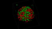
Image data courtesy of:
Dr. Yuji Mishima, Dr. Kiyohiko Hatake
Clinical Chemotherapy, Cancer Chemotherapy Center, Japanese Foundation for Cancer Research
Behaviors of Migration and Cell-cell Connection of Fibroblasts in A Multi-layered Myoblast Sheet
|
Image data courtesy of:
Eiji Nagamori, Ph.D Masahiro Kino-oka, Ph.D. Department of Biotechnology, Graduate School of Engineering, Osaka University |
Visualizing Retinoic Acid Signaling in A Zebrafish Embryo Using YFP as a Reporter
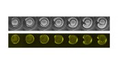
Image data courtesy of:
Satoshi Shimozono, Ph.D. Atsushi Miyawaki, M.D., Ph.D.
Laboratory for Cell Function Dynamics, Advanced Technology Development Core, RIKEN Brain Science Institute
Reference:
Shimozono S. et al. Visualization of an endogenous retinoic acid gradient across embryonic development. Nature 496, 363-366 (18 April 2013)
Observation of Antibody Dependent Cellular Cytotoxicity (ADCC)
|
RPMI 4788 cells (human colon cancer cell line) were treated with an antibody drug, cetuximab, and co-cultured with natural killer (NK) cells ADCC was observed using the FV10i after addition of NK cells
Cetuximab: Alexa Fluor 647 (red) NK cells: ZsGreen (green) Detection of dead cells: DAPI (blue) |
Image data courtesy of:
Dr. Yuji Mishima, Dr. Kiyohiko Hatake
Clinical Chemotherapy Department, The Cancer Chemotherapy Center of the Japanese Foundation for Cancer Research
Observation of the Effect of Anticancer Cisplatin with Different Concentrations
| Cell-cycle of HT-29 cells treated anticancer drug ,Cisplatin, was observed using time-lapse imaging of HT-29 expressing Fucci (a fluorescent cell-cycle indicator). The cells was treated by Cisplatin with different concentrations, control( 0 ug/ml), low(0.25 ug/ml), high(2.5 ug/ml) in 35 mm glass bottom dish after culturing for 48 hour. |
Image data courtesy of:
Dr. Yuji Mishima, Dr. Kiyohiko Hatake
Clinical Chemotherapy Department, The Cancer Chemotherapy Center of the Japanese Foundation for Cancer Research
Localization of Phosphoinositides During Cell–cell Fusion with Long Projections
| RAW264.7 cells expressing the reporter constructs in the presence of 1 µg/ml doxycycline were stimulated with 10 ng/ml RANKL for 48 h. Localization of PtdIns(4,5)P2 was visualized in red, while that of PtdIns(3,4,5)P3 was visualized in green. Frames were taken every 2 min for 34 min. Bar, 25 µm. |
Image data courtesy of:
Tsukasa Oikawa, Ph.D.
Department of Molecular Biology, Hokkaido University Graduate School of Medicine
Reference:
Oikawa T, et al. Tks5-dependent formation of circumferential podosomes/invadopodia mediates cell-cell fusion. J Cell Biol. 197(4):553-568(2012).
HeLa Cell*1
|
Dye: YFP (Actin)
Lens: 60XW NA1.2 Laser wavelengths: 473nm Time Interval: Every 3.5 minutes (TOTAL 12h) |
Mouse Brain
| Mouse brain fluorescently labeled with DAPI (nuclei - blue), Alexa 488 (Actin - green), Alexa 568 (Neurofilament - red). Image is comprised of 9 different regions of interest automatically acquired over 14 Z sections at 1024x1024 and stitched together using FV10i software. |
HeLa Cell*1
|
Time Interval: Every 20 minutes during a night
Green: GFP Magenta: Mito Tracker Red |
*1 Although it became one of the most important cell lines in medical research, it’s imperative that we recognize Henrietta Lacks’ contribution to science happened without her consent. This injustice, while leading to key discoveries in immunology, infectious disease, and cancer, also raised important conversations about privacy, ethics, and consent in medicine.
To learn more about the life of Henrietta Lacks and her contribution to modern medicine, click here.
http://henriettalacksfoundation.org/