Features
High-Resolution Deep Imaging without Sample Damage
Microscope Frames
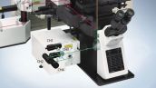
Upright Frame

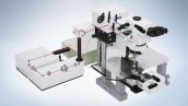
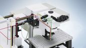
The FV1200MPE upright frame solution is dedicated to perform multiphoton microscopy task. This frame allows the end users to craft an optimal system during experiments are being performed that require synchronization of laser light stimulation and patch clamp signals. The dedicated high NA condenser detects transmitted second-harmonic generation (SHG) signals.
Learn more about upright frame
Inverted Frame
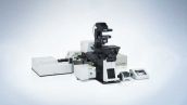
The inverted frame is ideal for the time lapse observation of thick, living specimens such as tissue cultures, and three-dimensional cell cultures. The inverted frame system also finds utility in intravital time lapse observation of organs and tissues through a body window.
Learn more about inverted frame
Laser and Scanner Systems
Brighter and Deeper Imaging with Less Damage
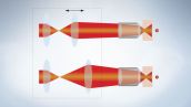
In multiphoton microscopy, fluorescence excitation efficiency is maximized by using a short pulse width in the focal plane. However, the pulse width of a femtosecond laser disperses as it passes through optics, broadening the pulse width when the beam exits from an objective. The laser beam-shaping optics establish a compensatory dispersion, the exact inverse of that produced by the microscope's optics (negative chirp), thus restoring the ideal pulse width for the specimen.
Custom Light Adjustment for the Exiting Laser Beam
The FV1200MPE is equipped with an AOM to adjust laser light. The AOM allows changes in laser intensity and rapid ON/OFF switching of the laser with microsecond control. This allows the laser output control to restrict irradiation to the region of interest, avoiding surrounding areas. In thick specimens, laser intensity and PMT voltages can be adjusted with specimen depth, allowing image capture without changes in image brightness.
Laser Beam Auto-adjust Capability
To achieve efficient multiphoton excitation, the laser beam, described by a Gaussian distribution of intensity, must fill the pupil diameter as it enters the objective. The beam expander of the FV1200MPE automatically adjusts the beam diameter depending on the objective and excitation wavelength. This optimizes laser beam characteristics for multiphoton excitation microscopy.
New Mirror Design Improves Excitation Efficiency
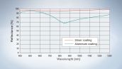
Our galvanometer mirror features an innovative silver coating that delivers outstanding reflective characteristics across a bandwidth from visible light to near-infrared. The total reflectivity rate of the XY scanner is also improved - providing as much as 25 % greater reflectivity in the near-infrared range compared to conventional aluminum mirrors. Additionally, where absolute power is essential, the increase in reflectivity translates into a 50 % improvement in multiphoton excitation efficiency compared to the FV1000MPE, making the mirror ideal for deep observation.
Detection Systems
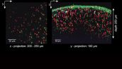
Sensitive Detection with Wide Field of View
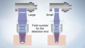
In multiphoton excitation, fluorescence is emitted from the focal spot inside the specimen. Cells and tissue components scatter light such that it emerges from the surface of the specimen at some distance from the incident beam. Incorporating a wide field of view, the FV1200MPE can capture the maximum amount of fluorescent signal, including scattered light, to provide highly efficient fluorescence imaging in scattering tissue.
Sensitive GaAsP Detector for Upright Microscope
Achieve images with a high S/N ratio, even in cases of extremely faint fluorescence, with a detector that makes use of hand-selected gallium arsenide phosphide (GaAsP, with 45 % QE).

Reflected Fluorescence Light Detector
Fluorescent signals are not only extremely faint, but also scatter within a thick specimen, causing further decay in signal intensity. The FV1200MPE uses a detector installed at a position close to the specimen in order to maximize detection efficiency.
Learn more about four channel
Learn more about two channel
Transmitted Fluorescence Light Detector
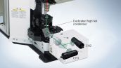
A high NA condenser and transmitted fluorescence light detector for multiphoton imaging detect fluorescence emitted from the focal plane and light scattered within the specimen. With this transmitted light detector, fluorescence can be detected with a high level of efficiency, especially in deep layers of the specimen.

Dedicated Objectives for Multiphoton Microscopy
Corrected Light Diffraction Delivers Deep Imaging
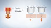
Refraction index differences within the specimen create a problem in deep imaging by disrupting the focal spot. The FV1200MPE’s dedicated objectives compensate for the refractive index mismatches thanks to its correction collar, allowing the formation of an ideal focal spot deep within the specimen without loss of energy density.
Take Imaging to New Depths
Olympus makes it possible to perform high-precision imaging of transparent biological specimens at exceptionally deep tissue levels with an innovative solution. This comprises of a dedicated 4 mm working distance objective for multiphoton imaging and a groundbreaking aqueous agent that renders biological specimens transparent.
Learn more Multiphoton Objectives
Learn more Optical Clearing Agent SCALEVIEW-A2
Powerful Optimization for High-Speed Imaging
Stimulation Systems
Simultaneous Multiphoton Imaging and Stimulation
Laser light stimulation can be adjusted as desired without the user being limited by imaging settings. This is due to the independent FV1200’s second scanner (SIM) used for laser light stimulation (available as an option). Connected to the SIM-scanner, the second multiphoton laser provides simultaneous stimulation at the same focal plane that is used for imaging.
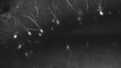
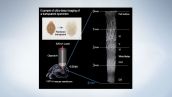
Calcium signal of a single dendritic spine examined by multiphoton uncaging and fluorescence
At the head of the single spine (red), multiphoton uncaging of caged glutamate was done and glutamate was injected (excitation of 720 nm). A line scan was performed on the line (the line linking the 2 triangles) from the head of this single spine toward the dendritic trunk. Calcium flow into the trunk via NMDA receptors at the head of the spine is apparent from these observations.
Reprinted from Noguchi et al. Neuron 46(2005)609-622.
Jun Noguchi, Haruo Kasai
Center for Disease Biology and Integrative Medicine, Faculty of Medicine,
University of Tokyo.
Multiphoton and Visible Light Stimulation
Multiple point stimulation software (optional) allows continued stimulation switching between IR and visible in one experiment. For example, uncaging with multiphoton excitation followed by channel-rhodopsins visible light stimulation without the need to stop image acquisition.
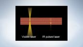
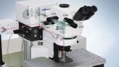
Example of integrated DM for light stimulation
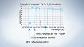
For example, with the dichromatic mirror indicated below, stimulation can be done with visible light at 488 nm and 559 nm; excitation can then be done with IR light at 920 nm to allow observation.
Versatile Imaging and Measurements
Wide Choice of Scan Modes

The FV1200MPE comes with AOM as standard and provides fine position and time control of imaging and light stimulation. Using Olympus tornado scanning allows rapid bleaching and laser light stimulation of desired fields in experiments like those involving FRAP and uncaging.
High Speed Optogenetics and Uncaging
Standard raster scanning modes are not capable of measuring high speed physiology with high signal-to-noise. Olympus’ innovative Multi point Mapping Advanced Software (MMASW) provides a solution for the most demanding high speed measurements. Using methods similar to Random Access Scanning and Targeted Path Scanning, users have the freedom to choose their own rapid multi point measurement or stimulation paths. Each scanned position can be expanded to a larger area. Silver galvanometer
mirrors drive the scan, ensuring access to full field of view and multicolor scanning capability with excellent light throughput for both visible and IR lasers. Measure cell network fluctuations at up to 101 cycles per second, with up to 50,000 Hz data output per position. High signal-to-noise captures all the data you need.
Use the Mapping feature to quickly identify fluorescence fluctuation positions or areas of high signal and automatically assign positions for high speed measurements.
Map cellular responses to simultaneous stimulus with Olympus’ unique SIM scanner for optogenetics or uncaging experiments (FV1200MPE BASIC and TWIN only).
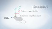
Synchronize detection with electrophysiology or other external devices. High speed electrophysiology, calcium measurements, optogenetics and FRAP/FLIP experiments. SIM scanner stimulation synchronized with imaging ensures cell responses during or immediately following stimulation are captured as they happen. The stimulation/imaging positions and laser wavelengths can be set separately with two independent beams.
Synchronized Laser Stimulation and Patch Clamping
The FV1200MPE's analog unit enables voltages to be converted into images and handled just like fluorescence images. For example, electrical signals measured by patch clamping during laser light stimulation can be synchronized with the image acquisition and displayed with pseudo-color.
Electrophysiology and Rapid Measurements
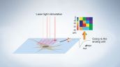
The observation field is divided into a grid and separate fields are discretely irradiated with a laser, allowing laser light stimulation while excluding the signal influence from adjacent fields. The mapping & multi point software enables auto stimulation at multiple points (optional software).
Brightness Compensation in the Z Plane
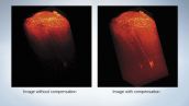
Sample brightness typically decreases when imaging deeper into a thick specimen. Use of this function enables changing of the detector sensitivity and laser power while continuously acquiring an image to match the focal position, thus allowing high-sensitivity and high-precision imaging without losing information from the thick portion of the specimen.
Arm Height Raising Kit for Small Animal Work
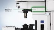
The arm height raising kit provides an additional 40mm of clearance and is mounted between the microscope frame and the reflected light illuminator. This facilitates experiments requiring small animals.
Multiple Options for System Configuration
M System: Multiphoton Exclusive with M Scanner
This multiphoton exclusive system is not equipped with visible light lasers. Simple optics optimized for multiphoton microscopy allow a smaller size, simpler operation, and deeper imaging within the specimen. The system uses a gold-coated galvanometer scanning mirror.
B System: Multiphoton with Standard Scanner
This system is equipped with an IR laser for multiphoton imaging and laser for visible light, so it is designed for deep imaging by multiphoton microscopy and confocal imaging with a visible laser. The system is designed for a variety of imaging including Live Cell and in vivo imaging. Using this system along with the double laser combiner allows multiphoton imaging and visible light stimulation.
S System: Multiphoton Laser Light Stimulation

This system is equipped with an IR laser delivering the light to the scanner for stimulation. In addition to general multiphoton microscopy, the system allows pinpoint light stimulation by multiphoton excitation during imaging with a visible laser. Multiphoton microscopy does not allow some image acquisition modes such as Time Controller.
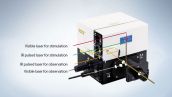
T System: Multiphoton Imaging and Stimulation
This system synchronizes laser light from 2 independent IR lasers for stimulation and imaging. It provides the multiphoton imaging capability of visualizing deep within the tissue, while at the same time, enabling pinpoint 3D stimulation with multiphoton excitation, for example, when stimulating a single dendritic spine located deep within the tissue. The newly introduced SIM dual port feature allows the SIM scanner to accurately stimulate with both visible laser as well as IR laser.
Laser Sharing System
This System Allows 2 Microscopes to Share a Single Laser. Example of a B system (Basic system) sharing a laser with an M system (Multiphoton exclusive system) Both the B system’s BX61WI and M system’s BX61WI share a single laser.
Visible Lasers for Standard Confocal Microscopy
The multi-combiner enables combinations with all of the following diode lasers: 405 nm, 440 nm, 473 nm, 559 nm and 635 nm. The system can also be equipped with conventional Multi-line Ar laser and HeNe-G laser.
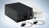
Double output type
The multi-combiner outputs laser light with two fibers. Light can be used for both observation and laser light stimulation.
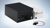
Single output type
The multi-combiner with a single output fiber for visible light observation. AOTF is standard equipment.
FLUOVIEW FV1200MPE femtoCARS Add-on
The femtoCARS equipped FV1200MPE provides a label-free lipid imaging capability based on molecular vibrations using the Coherent Anti-Stokes Raman Scattering (CARS) microscopy method.
Sorry, this page is not
available in your country.