Live cell dynamics at 3 fps and 120 nm resolution or 200 fps in standard resolution mode
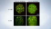
Olympus’ SD-OSR microscope achieves super resolution by combining superior optics with the new Yokogawa CSU-W1 Spinning Disk to improve signal and reduce noise. In super resolution mode, Airy disk oversampling drives resolution down to 120 nm by capturing subtle high-frequency spatial components not visible in conventional spinning disk microscopy. No special protocols are required; use familiar imaging workflows and achieve higher resolution results.
Superior live cell confocal imaging
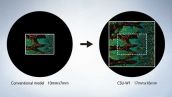
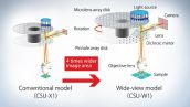
The new Yokogawa CSU-W1 incorporates significant advancements over its predecessors and is optimized to leverage the capabilities of sensitive SCMOS large format detectors while meeting the requirements of today’s live cell imaging. In addition to a larger disk and quadrupled field of view, the new disk features a reduced pinhole pitch which increases signal to noise ratios, improves image contrast, and increases the effective confocal working distance before signal crosstalk effects appear.
Superior Confocality and Reduced Crosstalk
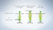
Larger Field of View
Olympus silicone oil objectives for high SNR
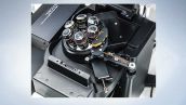
The Olympus 1.35 NA Silicone oil objective is an integral component of the system.
Correct objective selection is most important in order to obtain the most signal and highest resolution possible. By matching the refractive index of the sample to the immersion medium, silicone oil objectives minimize spherical aberration resulting in deeper, brighter, and higher resolution imaging. Olympus silicone oil objectives have high numerical apertures (NAs) and correction collars that enable users to optically compensate for spherical aberration, and use a silicone based oil which most closely matches the refractive index of living samples.
The importance of a correction collar
Spherical aberration is influenced by refractive index mismatches in the optical path, e.g. varying thickness of coverslips, observation depth of the sample, composition of cells or tissues, and changes in temperature. High NA objectives are particularly susceptible to these effects. Adjusting the correction collar of your objective is essential to compensate for spherical aberration to improve image quality.
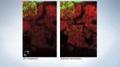
Comparison of oil and silicone immersion 60X objectives In a glycerol-mounted Drosophila brain.
Ease of access
The SD-OSR incorporates Olympus’ exclusive correction collar arm for easy access to the spherical aberration correction collar. This translates access to the correction collar to the right side of the nosepiece, eliminating the need to remove the sample in order to make adjustments. It’s now much easier to iteratively adjust the collar in combination with sample focus to minimize spherical aberration and optimize signal.
Optimized and simplified for live cell imaging
The design of the SD-OSR makes it an easy fit into any fluorescence imaging workflow. In contrast to competing localization and depletion mode approaches to super resolution, SD-OSR does not require any specific fluorochromes or sample conditions to obtain dynamic 120 nm resolution imaging of living systems. The SD-OSR is transparent to existing workflows and directly enhances existing microscopy with no change in sample preparation. The SD-OSR is simple to use and rapidly integrates into any imaging environment with minimal user training.
Three imaging modes in one system
The SD-OSR supports three distinct imaging modes: standard widefield, high performance live cell confocal, and live cell SIM to 120nm resolution. When placed into widefield "bypass" mode, the SD-OSR functions as a high quality IX83 inverted widefield microscope and fully supports 3D and multi-channel imaging. In spinning disk mode the system the Yokogawa CSU W1 provides superior optical contrast available at frame rates up to 200 frames/second. In SIM mode the SD-OSR produces resolution limited point scanning and well as optical oversampling to achieve 120nm planar resolution at up to three frames per second.
The flexible imaging capability of the SD-OSR provides that you’ll be able to obtain the best possible imaging of your sample, under any circumstances.
Designed for living systems
SD technology is proven for its ability to produce live cell confocal images of excellent quality. Yokogawa’s microlens and Nipkow disk technology balance speed, sensitivity, and resolution while minimizing cell stress. The system can be easily integrated to include stage top incubation and environmental control to further promote biological sample integrity data relevance. SD-OSR MetaMorph automation captures the living dynamics in three dimensions and multiple fluorescent channels over time.