Objectives play a critical role in your microscope system since they help to establish image quality. The reality is, many conventional objectives introduce imaging challenges that can compromise your microscope’s performance.
Fortunately, optical technology continues to advance and correct common imaging errors. Today’s blog post will explore three advantages of using Olympus’ high-performance X Line™ objectives to enable high-quality imaging data.
1. Expanded flatness for uniform quality.
Using conventional objectives, it can be challenging to achieve uniformly flat images in a large field of view (FOV). Poor image flatness can cause dark or blurry image edges, reducing image quality and making it difficult to analyze or measure samples.
How can our X Line objectives help? Our objectives provide outstanding image flatness from the center to the edge in large FOV observations. The consistently high image quality across the entire field of view helps you create precisely stitched images of large specimens, such as whole tissue sections. You can also rest assured that you have reliable image data for quantitative analysis.
Let’s look at an example:
 |  |
Comparing image flatness between conventional objectives (left) vs. X Line objectives (right).
These 12 × 12 images of a Fucci2 Tg mouse brain section were captured with the FV3000 confocal microscope using conventional and X Line 60x oil immersion objectives (NA 1.42). (Cyan: DAPI (405 nm).)
As you can see, the improved flatness of the X Line objective delivers a clearer tiled image in a wide FOV.
(Images courtesy of Takako Kogure and Atsushi Miyawaki from the Laboratory for Cell Function Dynamics, RIKEN Center for Brain Science.)
2. Enhanced chromatic correction for exceptional color reproducibility.
Chromatic aberration is a common optical challenge. It occurs when a lens fails to focus different colors in the same place. Essentially, the lens acts like a prism — colors that pass through the lens split at different angles. Unless corrected, microscopy images will look distorted with colored fringes around the edges. Despite this, many conventional lenses offer insufficient chromatic correction.
Our X Line objectives provide broad chromatic aberration correction from 400–1000 nm. This results in much better color reproducibility during brightfield and multicolor fluorescence imaging. Accurate color reproduction also improves image reliability, so you can feel confident in your data.
See the difference for yourself in the following images.
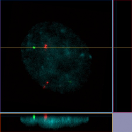 | 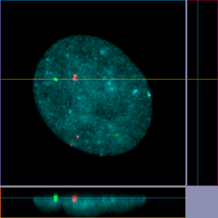 |
Comparing color reproducibility between conventional objectives (left) vs. X Line objectives (right).*
These images of a HeLa cell labeled using the FISH technique were captured with the FV3000 confocal microscope using conventional and X Line 60x oil immersion objectives (NA 1.42). (CEP17 (Spectrum Green), CEP18 (Spectrum Orange), and the nucleus (DAPI)).
With the X Line objective’s wide chromatic correction range of 400–1000 nm, green and red signals show up accurately within the nucleus. In the image taken with conventional objectives, signals located at the bottom of the cell nucleus appear outside it.
3. Improved numerical aperture for excellent image quality.
An objective’s numerical aperture defines its ability to gather light and determines the image resolution. Essentially, an objective with a higher numerical aperture can capture higher resolution, brighter images, so you can see more of your sample.
In contrast, an objective with high magnification but low numerical aperture will limit your ability to see sample details, increasing your risk of overlooking important information.
Our X Line objectives offer a higher numerical aperture up to 1.45 to help ensure excellent image quality for quantitative analysis. Improved numerical aperture can help with many applications, such as detecting dim signals during fluorescence observation or minimizing phototoxicity/photobleaching during fluorescence live cell imaging experiments.
In the example below, see how the higher NA of our X Line objectives improves the brightness.
 |  |
Comparing image resolution between conventional objectives (left, NA 0.75) vs. X Line objectives (right, NA 0.8).
These widefield fluorescence images of NG108-15 cells were captured using conventional and X Line 20x dry objectives. (Blue: nucleus, green: microtubules, red: actin filaments.)
As you can see in the image comparison, the improved numerical aperture of the X Line objective captured more light and led to a brighter image.
Excellent objectives enable efficient experiments
Conventional objectives often require you to make a tradeoff between these three important advantages—numerical aperture, image flatness, or chromatic correction. If you switch objectives to achieve an improvement in one area, it may compromise another.
Olympus’ X Line high-performance series are built with ultra-thin lenses to help you achieve all three benefits in one objective, enabling more efficient experiments. Learn more about the X Line series to find the right objectives for your application.
*Although it became one of the most important cell lines in medical research, it’s imperative that we recognize Henrietta Lacks’ contribution to science happened without her consent. This injustice, while leading to key discoveries in immunology, infectious disease, and cancer, also raised important conversations about privacy, ethics, and consent in medicine.
To learn more about the life of Henrietta Lacks and her contribution to modern medicine, click here.
http://henriettalacksfoundation.org/
Related Content
Video: X Line High-Performance Objectives Technical Introduction
Video: X Line High-Performance Objectives Product Introduction
White Paper: X Line Objectives Offer Revolutionary Optical Performance