Multiplexing with the FLUOVIEW FV3000 Confocal Microscope
Characterizing the Morphology of Mouse Medial Prefrontal Cortex
Studying cognitive impairment mechanisms requires the ability to associate morphological changes with physiological responses. To understand how the states and treatments of diseases relate to and affect brain morphology, it is important to be able to identify multiple morphological structures within the same sample. In this study, the FV3000 confocal microscope with TruSpectral detectors was used to successfully image six different structures in mouse medial prefrontal cortex (mPFC): astrocytes, pyramidal neurons, inhibitory neurons, neuronal membranes, axon initial segments, and nuclei.
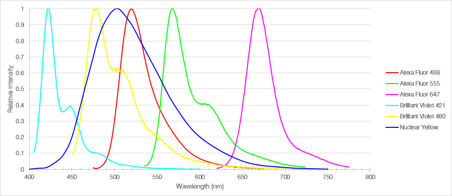
Figure 1: Emission spectra of the six fluorophores used to label mouse medial prefrontal cortex sections.
TruSpectral Technology Enables Distinct Identification of Six Structures
In this experiment, a 30 µm-thick fixed section of mouse medial prefrontal cortex was labeled with six different fluorophores. An Olympus FV3000 confocal microscope equipped with a UPLSAPO20X objective captured 3D fluorescent images of the multiplexed brain tissue. Using TruSpectral detection, individual acquisition and detection parameters were set for each fluorophore to optimize signal detection and prevent bleed-through. The resulting image produced bright signals of each fluorophore with distinct emission profiles, enabling accurate detection of multiple structures within the same sample.
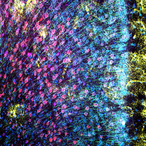
Figure 2: Mouse mPFC labeled with glial fibrillary acidic protein (GFAP; astrocyte marker; yellow), calmodulin-dependent protein kinase II (CaMKII; pyramidal neuron marker; red), amphoterin-induced protein 1 precursor (AMIGO-1; neuronal membrane marker; cyan), parvalbumin (PV; inhibitory neuron marker; purple), ankyrin-G (AnkG; axon initial segment marker; green), and nuclear yellow (nuclei marker; blue).
Imaging conditions
Microscope: FLUOVIEW FV3000 laser scanning confocal microscope (virtual channel mode)
Objective: 20X air objective (UPLSAPO20X)
Lasers: 405 nm (BV421, Nuclear Yellow), 445 nm (BV480), 488 nm (AF488), 561 nm (AF555), and 640 nm (AF647)
100X Silicone Oil Objective Enables High-Resolution Imaging of Distinct Morphological Structures
To obtain higher resolution images of the mouse mPFC, the 100X silicone oil objective was used with TruSpectral high-sensitvity detectors to obtain bright, detailed images of a region of the cortex exhibiting all six markers. This combination enabled researchers to clearly observe the morphology of the different population of neurons within the medial prefrontal cortex.
(A)
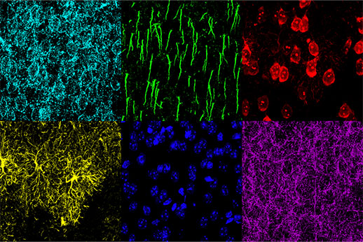 |
(B)
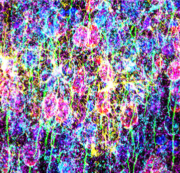 |
Figure 3: Mouse mPFC labeled with glial fibrillary acidic protein (GFAP; astrocyte marker; yellow), calmodulin-dependent protein kinase II (CaMKII; pyramidal neuron marker; red), amphoterin-induced protein 1 precursor (AMIGO-1; neuronal membrane marker; cyan), parvalbumin (PV; inhibitory neuron marker; purple), ankyrin-G (AnkG; axon initial segment marker; green), and nuclear yellow (nuclei marker; blue). (A) Individual channels of the six fluorophores. (B) Overlay image.
Imaging conditions
Microscope: FLUOVIEW FV3000 laser scanning confocal microscope (virtual channel mode)
Objective: 100X silicone oil objective (UPLSAPO100XS)
Lasers: 405 nm (BV421, Nuclear Yellow), 445 nm (BV480), 488 nm (AF488), 561 nm (AF555), and 640 nm (AF647)
How the FV3000 Confocal Microscope Facilitated Our Experiment
Fully Spectral System with High-Efficiency GaAsP Detectors Provides High Sensitivity for Multiplex Imaging
| Integrated in all FV3000 confocal microscopes, TruSpectral detection technology enables higher light throughput compared to conventional spectral detection units. The volume phase hologram diffracts light with up to three-fold higher transmission efficiency versus reflection gratings. With the FV3000 microscope’s high-sensitivity spectral detector (HSD), both bright and dim signals can be optimally separated by independently adjusting the sensitivity of each TruSpectral detector. Furthermore, the FV3000 system’s spectral deconvolution algorithm enables overlapping spectra to be separated based on the spectral information from lambda stack images. Fluorescence cross-talk between channels can be eliminated by the unmixing algorithm during both live image acquisition and post-processing, enabling clean separation of up to sixteen fluorophores. | Related Videos |
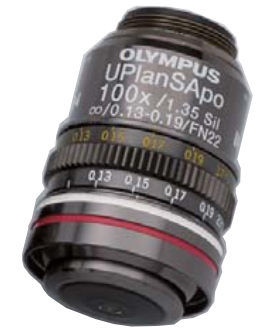
UPLSAPO100XS Silicone Oil Immersion Objective for Bright Imaging of Thick Tissue
The UPLSAPO super apochromat objectives compensate for both spherical and chromatic aberrations, and they have high transmission from the visible to the near-infrared region. The refractive index of silicone oil (ne≈1.40) is close to that of living tissue (ne≈1.38), enabling high-resolution observations deep inside living tissue with minimal spherical aberration and accurate 3D morphology.
|
Magnification: 100X
NA: 1.35 (silicone oil immersion) W.D.: 0.2 mm Correction Level of Chromatic Aberration: Super Apochromat (SAPO) |
Using the FV3000 Microscope to Research the Impacts of Neuropathic Pain on the Brain at the Molecular Level
Comment by Stephanie Shiers
 | Our work focuses on medial prefrontal cortex (mPFC)-dependent cognitive impairment in neuropathic pain. We investigate structural plasticity in the mPFC to elucidate the cell-types involved in cortical dysfunction and to gain insight into the functional changes of these cells. We have observed a reduction in the length of axon initial segments (AIS) in the mPFC of male mice with neuropathic pain that correlates to poor performance in a cognitive task (Shiers, et al. J Neurosci 2018, Shiers, et al. Neuropsychopharmacology, 2019). Understanding the morphology of these cell types provides us with clues to what underlies cognitive dysfunction in pain, and how we can treat it. High-resolution imaging with the FV3000 microscope has been critical for our analyses of the mPFC microarchitecture. |
Comment by Dr. Theodore Price
 | A primary goal of our research is to understand how neuropathic pain impacts brain structures that are intimately involved in cognitive processing. We know that cognitive problems are an important comorbidity of neuropathic pain, but we know little about the molecular underpinnings. Our work on the mouse PFC has revealed fine changes in the AIS that are caused by neuropathic pain. Our ability to obtain the highest quality images of the ultrastructure of the PFC on the FV3000 microscope has opened up new ways for us to understand this important aspect of neuropathic pain. |
Acknowledgments
This application note was prepared with the help of the following researchers:
Stephanie Shiers, Ph.D. Candidate, Price Lab, University of Texas at Dallas
Theodore J. Price, Ph.D., Price Lab, Eugene McDermott Professor, Director,
Center for Advanced Pain Studies, Department of Neurobiology, School of Behavioral and Brain Sciences, University of Texas at Dallas
Products related to this application
Sorry, this page is not
available in your country.