Analysis of Anticancer Drugs-Targeting the Interior of Spheroids
The analysis of fluorescent images combined with the use of the cell imaging analysis system enables the characterization of compounds targeting a hypoxic area inside spheroids.
Objectives
Cell heterogeneity in tumor tissue is closely related to cancer progression, angiogenesis, drug resistance, and other issues. Therefore, cell heterogeneity is becoming an important factor in drug development strategy. Since an in vitro cancer spheroid model can recapitulate a microenvironment of solid tumors in living tissue, it is expected to be a useful tool in analysis of heterogeneity in cancer cells. The cell imaging analysis system quantitatively analyzes image data obtained from confocal fluorescent observations. In this study, the cytotoxicity of a drug was quantitatively evaluated through the visualization of spheroid size and microenvironment inside spheroids.
Preparation of samples
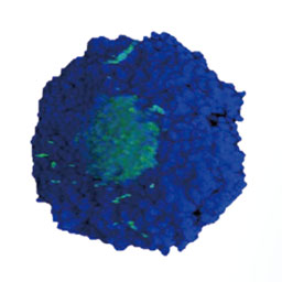
Cancer spheroids (TH-29) were prepared using a PrimeSurface® 96U plate (SUMITOMO BAKELITE CO., LTD). Hoechst 33342 and MAR probe (GORYO Chemical) were added to the spheroids one day before being measured by the system. The size and hypoxic area of the spheroids were then visualized.
Conclusion
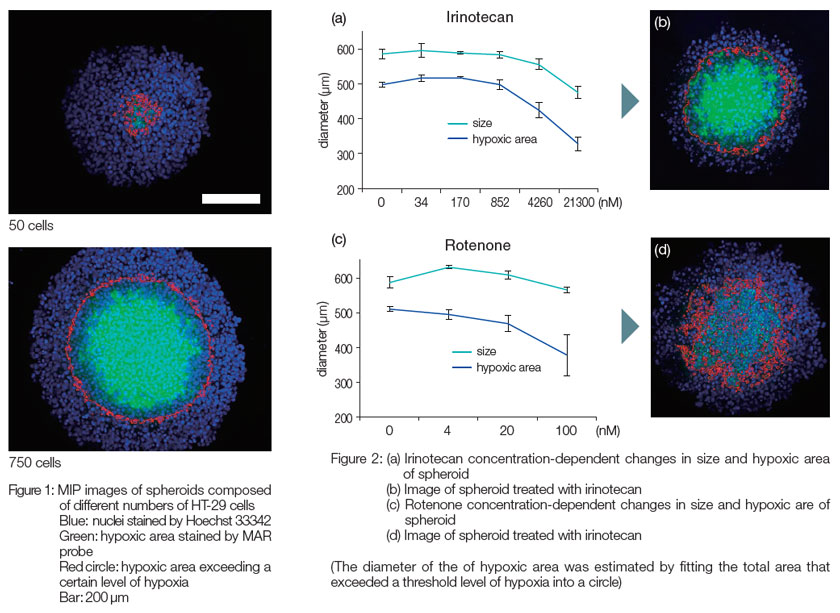
According to maximum intensity projection images of spheroids composed of different numbers of HT-29 cells, the hypoxic area increased with an increase in spheroid size (Figure 1). The treatment of spheroids that had a large hypoxic area with irinotecan (topoisomerase inhibitor) resulted in the simultaneous reduction in both spheroid size and hypoxic area (Figure 2(a) and (b)). However, treatment with rotenone (an inhibitor of the electron transfer chain in mitochondria) resulted only in a reduction in hypoxic area without a reduction in spheroid size (Figure 2(c) and (d)). These results indicated that the system enables the quantitative evaluation of different effects of drugs on heterogeneity in cancer cells.
PrimeSurface is a registered trademark of Sumitomo Bakelite Co., Ltd.
Olympus is a registered trademark, and NoviSight and Insightful Analysis, Intelligent Answers are trademarks of Olympus Corporation.
Products related to this application
Sorry, this page is not
available in your country.