Fluorescent Image Analysis–Live Dead Cell Assay of Spheroids
The ratio of dead cells inside spheroids and drug efficacy can be quantitatively evaluated using a confocal microscope and NoviSight™ software's cell counting module.
Objectives
The distance from the surface of a tumor to the core has a diffusion gradient of oxygen or nutrients, and this severe environment causes resting cells or dead cells deep inside the tumor tissue. A spheroid model is expected to recapitulate tumor microenvironment. Previously, each section had to be separately prepared to observe the inside of 3D samples. However, the development of tissue clearing technology now makes it possible to observe inside 3D samples while preserving their structural integrity. In this study, confocal images of fluorescent stained cancer spheroids, treated with a drug, fixed, and made transparent, were subjected to 3D quantitative analysis using NoviSight™ software. The study demonstrates that drug-independent and drug-dependent cell death can be confirmed using a clearing reagent and NoviSight™ software.
Preparation of samples
A cell suspension of HT-29 was seeded into PrimeSurface®96U plates (SUMITOMO BAKELITE CO., LTD), a U bottom well plate, at 500 cells/well. Staurosporine (STS) at various concentrations and NucView550 were added to each well 8 days after the start of cell culture. After incubation for 1 day, cells were fixed with 4% paraformaldehyde and permeabilized with 0.5% TritonX-100/PBS. Then, cell nuclei were stained with Hoechst 33342 overnight at 4 °C, and spheroids were transparentized with ScaleS.
Conclusion
Acquisition and analysis of fluorescent images
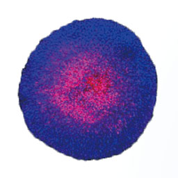
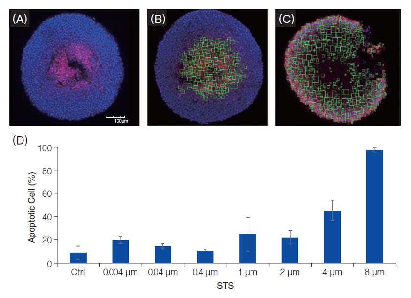
Confocal fluorescent images of the above-mentioned spheroids were obtained using the FV3000 laser confocal microscope. Even in the absence of STS, cell nuclei deep inside spheroids were stained with NucView550 (A), indicating that apoptosis takes place. Cell nuclei recognition and cell counting analysis using NoviSight™ software enable the quantification of the dead cell ratio (B), and we confirmed STS-induced cell death occurred in a dose dependent manner (C,D). These results demonstrate how NoviSight™ software can contribute to the quantitative evaluation of 3D cell death induced by drugs.
PrimeSurface is a registered trademark of Sumitomo Bakelite Co., Ltd.
Olympus is a registered trademark, and NoviSight and Insightful Analysis, Intelligent Answers are trademarks of Olympus Corporation.
Products related to this application
Sorry, this page is not
available in your country.