The Response of Mitochondria in Cancer Spheroids to Drugs
The effect of an uncoupling agent on mitochondria in spheroids can be visualized.
This is a promising method for testing toxicity to mitochondria.
Objectives
Mitochondria are indispensable organelles that supply cellular energy. Toxicity to mitochondrial function is an important factor to be tested for new compounds. In this study, the response of mitochondria in 3D cell cultures to drugs was investigated.
Preparation of samples
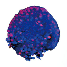
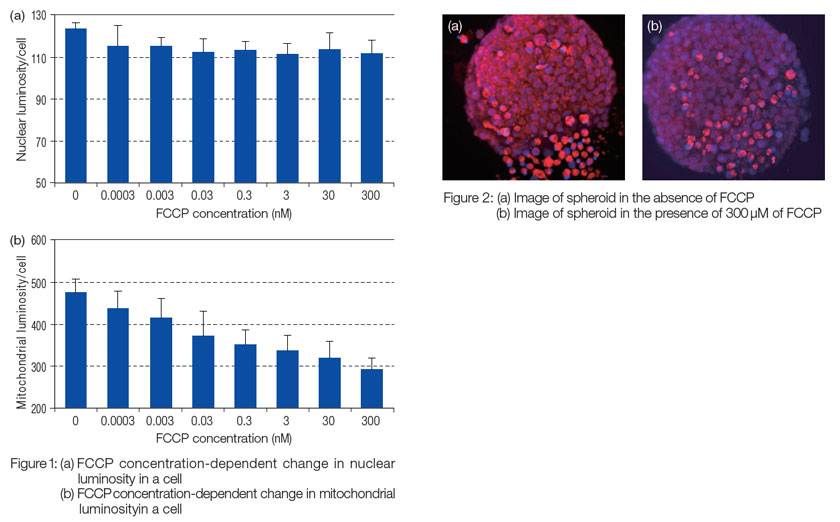
A cell suspension of HT-29 was seeded to a U-bottom 96-well plate and incubated for 96 hours to prepare 3D cell cultures. Spheroids were incubated with MitoTracker® Red CMXRos (a membrane potential-dependent, mitochondrial staining dye) and FCCP (uncoupling agent, 0-300 μM) for 45 minutes, followed by fixing in 4% formalin and nuclear staining with DAPI.
Conclusion
Acquisition and analysis of images
Confocal fluorescent images of the above-mentioned spheroids were obtained. The average luminosity of nuclei and mitochondria in a cell was estimated by superimposing images obtained every 5 μM using the mean intensity projection method. No effect of FCCP was observed on nuclei (Figure 1 (a)), while the average luminosity of mitochondria in a cell decreased with an increase in FCCP concentration (Figures 1 (b) and 2).
MitoTracker is a registered trademark of Molecular Probes, Inc.
Olympus is a registered trademark, and NoviSight and Insightful Analysis, Intelligent Answers are trademarks of Olympus Corporation.
Products related to this application
Sorry, this page is not
available in your country.