Evaluation of Drug Efficacy–The Response of Spheroids to a Drug
Confocal observation combined with the use of the system enables the evaluation ofdrug efficacy in spheroids stained with an appropriate fluorescent dye.
Objectives
The majority of efficacy assays of anticancer drugs have relied on the use of immobilized cells grown as 2D monolayers. However, these 2D monolayers are unable to recapitulate the complex native environment of cancer cells, such as 3D structure. Therefore, 3D cell cultures like spheroids are attracting attention as models that have the potential to improve the physiological relevance of cell-based studies. In this study, the efficacy of Staurosporine (STS) in spheroids was evaluated through confocal observation combined with the use of the cell imaging analysis system.
Preparation of samples
A cell suspension of HeLa was seeded into a PrimeSurface® 96U plate (SUMITOMO BAKELITE CO., LTD) at 500 cells/well. Serially-diluted STS was added to each well 48 hours after the start of cell culture. Hoechst 33342 (nuclear staining dye) and propidium iodide (PI: dead cell staining dye) were added to each well 24 hours after the addition of STS.
Conclusion
Acquisition and analysis of fluorescent images
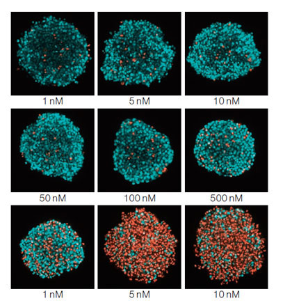
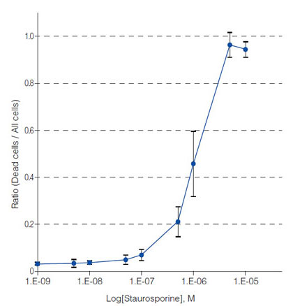
Fluorescent images of the above mentioned spheroids stained by Hoechst 33342 and PI were obtained using the system. As shown in Figure 1*, the ratio of dead cells (pseudo red) to all cells (pseudo blue) in each well increased with an increase in STS concentration. The ratios can be graphically displayed against STS concentration (Figure 2, bars: SD, n=8), using the nuclei identification and counting results generated by the analysis software. These results indicated that the system enables easy evaluation of drug efficacy in one microplate.
Figure 1: STS concentration-dependent dead cells/all cells images of spheroids* |
Figure 2: STS concentration-dependent dead cells/all cells ratio in spheroids |
PrimeSurface is a registered trademark of Sumitomo Bakelite Co., Ltd.
Olympus is a registered trademark, and NoviSight and Insightful Analysis, Intelligent Answers are trademarks of Olympus Corporation.
*Although it became one of the most important cell lines in medical research, it’s imperative that we recognize Henrietta Lacks’ contribution to science happened without her consent. This injustice, while leading to key discoveries in immunology, infectious disease, and cancer, also raised important conversations about privacy, ethics, and consent in medicine.
To learn more about the life of Henrietta Lacks and her contribution to modern medicine, click here.
http://henriettalacksfoundation.org/
Products related to this application
Sorry, this page is not
available in your country.