Evaluation of Drug Efficacy–The Response of Co-Cultured Spheroids to Drugs
Confocal observation combined with the use of the system enables the evaluation of drug efficacy in co-cultured spheroids stained with an appropriate fluorescent dye.
Objectives
The majority of efficacy assays of anticancer drugs have relied on the use of immobilized cells grown as 2D monolayers. However, these 2D monolayers are unable to recapitulate the complex native environment of cancer cells, such as those in a 3D structure. Therefore, 3D cell cultures like spheroids are attracting attention. Furthermore, co-cultured spheroid systems composed of two or more types of cells has proven to be useful in the study of intercellular interaction or cell activation mechanisms. In this study, the effect of chloroquine (CHL) on cocultured spheroids composed of U20S and HT-29 was investigated through fluorescent image analysis with the system.
Preparation of samples
A cell suspension containing YFP expressing transgenic U20S and HT-29 was seeded into a PrimeSurface® 96U plate (SUMITOMO BAKELITE CO., LTD) at 50 transgenic U20S cells/well and 450 HT-29 cells/well. Seriallydiluted CHL was added to each well 48 hours after the start of cell culture. Hoechst 33342 (nuclear staining dye) and propidium iodide (PI: dead cell staining dye) were added to each well 48 hours after the addition of CHL. The observation of YFP fluorescence enables U20S and HT-29 to be distinguished.
Conclusion
Acquisition and analysis of fluorescent images
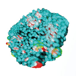
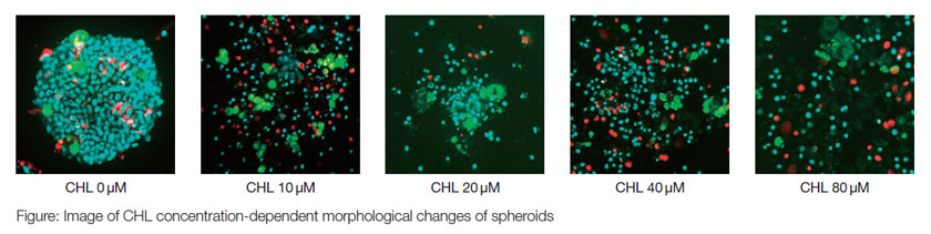
Fluorescent images of the above mentioned spheroids were obtained. As shown in the Figures, the co-cultured spheroids lost their 3D structural integrity of 3D structure with an increase in the CHL concentration. Interestingly, the HT-29 cells dispersed completely, while the U20S cells (pseudo green) remained aggregated. Also, the ratio of dead cells (pseudo red) to all cells (pseudo blue) in each well increased with an increase in CHL concentration. The ratios can be graphically displayed against CHL concentration, using the results from identification and counting of nuclei with analysis software. These results indicated that drug efficacy is easily evaluated using co-cultured spheroids in one microplate.
PrimeSurface is a registered trademark of Sumitomo Bakelite Co., Ltd.
Olympus is a registered trademark, and NoviSight and Insightful Analysis, Intelligent Answers are trademarks of Olympus Corporation.
Products related to this application
Sorry, this page is not
available in your country.