Evident LogoOlympus Logo
Anwendungsbeispiele

TIRF Imaging of Changes in Membrane Morphology and Molecular Dynamics


Total Internal Reflection Fluorescence (TIRF) Imaging of Changes in Membrane Morphology and Molecular Dynamics under the Cell Membrane with Olympus’ Z-drift Compensation System

Introduction

One important issue in current cell biology research is to understand the mechanism of physiological phenomena associated with the intercellular communication between adjacent cells. A promising step toward this goal is live cell microscopy that enables researchers to monitor changes in cell membrane morphology and the dynamics of localized molecules at the intercellular adhesion site. Figure 1 illustrates how high-precision TIRF imaging is enabling new types of advanced cellular research. The images, captured using an Olympus motorized inverted microscope IX series, show changes in the membrane morphology and molecular dynamics under the cell membrane. The Olympus Z-drift compensator maintained a sharp focus on the cells over a long period of time enabling these images to be captured in such high quality. This process demonstrates the importance of TIRF and the Olympus Z-drift compensator to advanced live cell imaging.

Time-lapse images of a Cos-1 cell
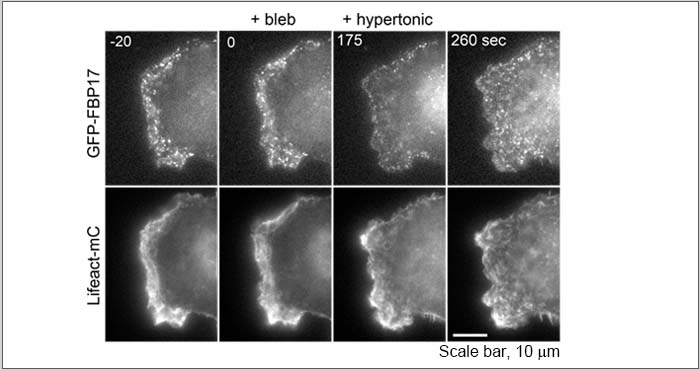
 Figure 1. Time-lapse images of a Cos-1 cell co-expressing GFP-17 and Lifeact-mCherry.

Examination of whether the recruitment of FBP17 to the plasma membrane is dependent on transient reduction of membrane tension caused by myosin based contraction force. FBP17 acutely disappeared from the cell edge after treatment with the myosin inhibitor blebbistatin (175 sec). This ef fect can be rescued by subsequent reduction of membrane tension induced by hypertonic buffer (260 sec), indicating that the FBP17 senses the membrane tension to assemble at the plasma membrane.

Time-lapse movie of a Cos-1 cell co-expressing GFP-17 and Lifeact-mCherry.

Imaging System;
Microscope: Research Inverted Microscope IX81
Objective: PlanApo 100XOTIRFM(100X, N.A.1.45)
CCD camera: Cascade II cooled CCD camera (Photometrics)
Z-drift Compensation System: IX-ZDC

Image data courtesy of;
Kazuya Tsujita, Ph.D., Toshiki Itoh, Ph.D.
Biosignal Research Center, Organization of Advanced Science and Technology, Kobe University

Reference;
Nat Cell Biol. 2015 Jun;17(6):749-58. doi: 10.1038/ncb3162.
J Cell Sci. 2013 May 15;126(Pt 10):2267-78. doi: 10.1242/jcs.12251

Polarisation of FBP17 is induced by PM tension increase.
COS-1cell co-expressing GFP-FBP17 and Lifeact-mCherry was observed by time-lapse microscopy upon hypotonic buffer. The movie was taken at 1 frame per 5 seconds and played at 15 fps.

Polarisation of FBP17 is disrupted by PM tension decrease.
COS-1 cell co-expressing GFP-FBP17and Lifeact-mCherry was observed by time-lapse microscopy upon addition of hypertonic buffer. The movie was taken at 1 frame per 5 seconds and played at 15 fps.

Polarisation of FBP17 is induced by PtdIns(4,5)P2 liberation.
COS-1 cell co-expressing GFP-FBP17, CFP-FKBP-PLC δ1 PH domain, and mRFP-FRB-MoA was observed by time-lapse microscopy upon addition of rapamycin. The movie was taken at 1 frame per 5 seconds and played at 15 fps.

Polarisation of FBP17 is disrupted by PtdIns(4,5)P2 depletion.
COS-1 cell co-expressing GFP-FBP17, CFP-PM-anchored FRB domain, and mRFP-FKBP-5-phosphatase domain was observed by time-lapse microscopy upon addition of rapamycin. The movie was taken at 1 frame per 5 seconds and played at 15 fps.

Dynamics of FBP17 at the leading edge. COS-1 cell co-expressing GFP-FBP17and Lifeact-mCherry was observed by time-lapse microscopy. The movie was taken at 1 frame per 5 seconds and played at 15 fps.

Acute disruption of FBP17 polarity by N-WASP inhibition. COS-1 cell co-expressing GFP-FBP17 and Lifeact-mCherry was observed by time-lapse microscopy upon addition of wiskostatin. The movie was taken at 1 frame per 10 seconds and played at 15 fps.

Acute disruption of FBP17 polarity by Arp2/3 complex inhibition.
COS-1 cell co-expressing GFP-FBP17 and Lifeact-mCherry was observed by time-lapse microscopy upon addition of CK-666. The movie was taken at 1 frame per 10 seconds and played at 15 fps.

Conclusion

Olympus’ live cell imaging solutions and Z-drift compensator facilitate long-term imaging studies of cellular processes. The Z-drift compensator utilizes low phototoxicity infrared (IR) light to detect the correct focus position, to make automatic focal adjustments, and to maintain precise focusing over time by avoiding focus drift due to factors such as temperature changes. The type of experiment described above cannot be accomplished using conventional microscopy because the images captured over time would be out of focus because of focus drift. The Z-drift compensator enables images to be captured without loss of focus. This facilitates chronological, high-precision tracking of dynamic changes of FBP17 and the Lifeact actin marker under the cell membrane.

Verwendete Produkte

TIRF-Mikroskopsysteme

IXplore TIRF

Das IXplore TIRF Mikroskopsystem wurde für Membrandynamik-, Einzelmoleküldetektions- und Kolokalisationsexperimente entwickelt und ermöglicht die simultane mehrfarbige TIRF-Bildgebung für bis zu 4 Farben. Das cellTIRF System von Olympus verfügt über eine stabile motorgesteuerte Steuerung für einen einzelnen Laserwinkel, die eine gleichmäßige Durchdringung mit evaneszenten Wellen für kontrastreiche, rauscharme Bilder ermöglicht. Unsere TIRF-Objektive zeichnen sich durch ein hohes SNR, eine hohe NA und Korrekturringe zur Anpassung an die Deckglasdicke und Temperatur aus.

  • Exakte Kolokalisierung von bis zu vier Markern durch separate Steuerung der Eindringtiefe
  • Nutzen Sie die Vorteile der TIRF-Objektive von Olympus mit der weltweit höchsten NA von 1,7*
  • Intuitive Planung komplexer Experimente mit dem Graphical Experiment Manager (GEM), cellFRAP und U-RTCE
* Stand 25. Juli 2017. Nach Angaben der Olympus Forschung.
Hochauflösende Objektive für Superauflösung/TIRF

APON-TIRF/UAPON-TIRF/UPLAPO-HR

Diese Apochromat-Objektive verfügen über unsere höchsten numerischen Aperturwerte und sind für eine kontrastreiche TIRF- und superauflösende Bildgebung optimiert. Mit der hohen NA der UPLAPO-HR-Objektive wird eine umfassende Planität erreicht, die eine hochauflösende Abbildung von Lebendzellen und Mikroorganellen in Echtzeit ermöglicht.

  • Hohe NA zur Erzeugung evaneszenter Wellen für kontrastreiche TIRF-Bilder oder Superauflösung
  • Die Objektive der HR-Serie sind die weltweit ersten* Plan-Apochromate mit NA 1,5 und hoher Verzeichnungsfreiheit

* Ab November 2018. Nach Angaben der Olympus Forschung.

Z-Drift-Kompensator

IX3-ZDC2

  • Stets im Fokus
  • Anwenderfreundliches Design
  • Speziell für das Lebendzell-Imaging
  • Hochpräzises Imaging mehrerer Bereiche mit der cellSens Software

wurde erfolgreich zu Ihren Lesezeichen hinzugefügt

Lesezeichen anzeigenSchließen

Maximum Compare Limit of 5 Items

Please adjust your selection to be no more than 5 items to compare at once

Sorry, this page is not
available in your country.

Sorry, this page is not available in your country