The Olympus Microscopy Resource Center galleries include images of fluorescent specimens, as well as darkfield, phase contrast, and Hoffman modulation contrast photomicrographs. In addition, the gallery features streaming video and images from featured microscopists.
Abramowitz
Mortimer Abramowitz, a renowned microscopist, received the New York Microscopical Society (NYMS) Ernst Abbe Memorial Award in 2002.
Olympus Fluorescence Gallery
Incident light fluorescence microscopy is rapidly growing as an investigational tool in the fields of medical and biological research.
Fluorescence Microscopy
Specimens featured in the fluorescence gallery are derived from a combination of stained thin sections, whole mounts, suspensions, and smears.
DIC
Thin unstained, transparent specimens are excellent candidates for imaging with classical differential interference (DIC) microscopy techniques.
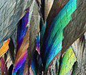
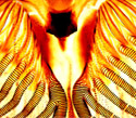
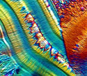
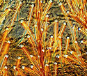
Polarized Light
Polarized light microscopy is a contrast-enhancing technique that may be utilized for quantitative and qualitative analysis of optically anisotropic specimens.
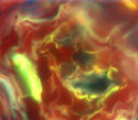
Darkfield Microscopy
Darkfield Illumination photomicrography contains a wide spectrum of images taken under a variety of conditions and utilizing many different specimens.
Hoffman Modulation Contrast Microscopy
Gallery of Hoffman Modulation Contrast photomicrography contains images taken under a wide variety of conditions using many different specimens.
Phase Contrast
Phase Contrast photomicrography contains a large collection of images taken under a wide variety of conditions using specimens.
Brightfield
Brightfield illumination has been one of the most widely used observation modes in optical microscopy for the past 300 years.
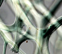
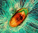
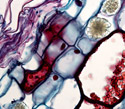
Plant Tissue
Autofluorescence in plant tissues is a phenomenon arising from endogenous biomolecules that absorb light in regions of the near-ultraviolet and visible light spectrum.
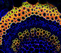
Rat Brain
The rat brain has served as an excellent model for elucidating the complex anatomy and physiological mechanisms of the human brain.
Fluorescence Digital Image Galleries
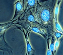
Cells in Culture
Many galleries displayed include images of fluorescent specimens, as well as darkfield, phase contrast, and Hoffman modulation contrast photomicrographs.
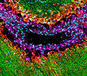
African Green Monkey
Many galleries displayed include images of fluorescent specimens, as well as darkfield, phase contrast, and Hoffman modulation contrast photomicrographs.
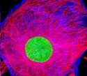
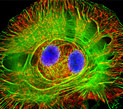
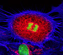
Fibroblast Cells
Many galleries displayed include images of fluorescent specimens, as well as darkfield, phase contrast, and Hoffman modulation contrast photomicrographs.
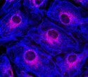
Epithelial Cells
Many galleries displayed include images of fluorescent specimens, as well as darkfield, phase contrast, and Hoffman modulation contrast photomicrographs.
Confocal Microscopy Digital Image Galleries
Cells in Culture
Galleries displayed include images of fluorescent specimens, as well as darkfield, phase contrast, and Hoffman modulation contrast photomicrographs.
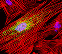
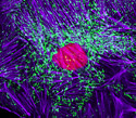
Endothelial Cells
Galleries displayed include images of fluorescent specimens, as well as darkfield, phase contrast, and Hoffman modulation contrast photomicrographs.
Epithelial Cells
Galleries displayed include images of fluorescent specimens, as well as darkfield, phase contrast, and Hoffman modulation contrast photomicrographs.
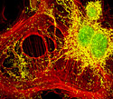
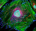
Fibroblast Cells
Galleries displayed include images of fluorescent specimens, as well as darkfield, phase contrast, and Hoffman modulation contrast photomicrographs.