
As research institutions limit time spent in the lab to practice social distancing and help keep staff safe, having the ability to automate and streamline tasks through live cell imaging is more important than ever to stay productive.
Yet, there are many considerations to keep in mind to optimize your equipment and avoid unexpected scenarios that might require re-entry to the lab.
Here are 4 tips to consider:
1. Keep samples healthy in a warm, stable environment.
One of the biggest challenges during live cell imaging is how to keep your samples healthy and behaving normally on the microscope stage—outside of their warm and comfortable incubator.
While this can be tricky, you can keep your samples as comfortable as possible by using microscope-dedicated stage-top incubators or enclosed incubators that can provide a stable temperature, humidity, and CO2 density for cells.
And don’t forget to heat your objective—this is a common source of cold contact to the samples that can harm their health.
Also remember that the peripheral region of microplates might have a higher risk of environmental fluctuation than the central region, so be sure to use the central wells and avoid the wells around the edge to improve stability. For instance, if you’re using a 96-well plate (12×8 wells), then only use the central 60 wells (10×6 wells) for the experiment.
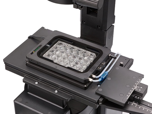
Figure 1. Stage-top incubator.
2. Create a stable environmental temperature to improve focus.
Another factor to consider is the temperature in the room, as a small change can affect imaging performance.
Environmental temperature stability for your microscope is especially critical for high magnification observation of live cells. Since high-NA observation has a shallow field depth, your system can lose focus even with a small Z-drift caused by temperature change.
To optimize the environment, make sure that your air conditioner is working and the room temperature is stable before starting the experiment. Also, avoid direct air flow from the AC to the microscope to improve stability.
3. Precisely set your focus and correction collar to enhance image quality.
Another way to address temporal fluctuation like Z-drift is to begin the live cell experiment by setting your focus and collar correction as precisely as possible.
Often researchers will adjust the focus carefully but forget about the correction collar. Yet, the correction collar can significantly improve image quality, particularly during deep tissue observation or when using a high NA objective.
The ideal position of the correction collar depends on many factors, such as the refractive index of the sample, the depth of the observation plane, and the thickness of the cover glass. Even though most glass cover slip and glass bottom dishes have a thickness of 170 µm (#1.5), it can fluctuate, and more importantly, we can’t assume these values with plastic vessels.
For this reason, always adjust the correction collar precisely by checking the image contrast, particularly when using plastic microplates or dishes.
Typically, collar correction must be done on site. For experiments that need proactive collar correction adjustments, such as deep observation with a multiphoton microscope, this can mean more time in the lab. The good news is that you can automate these adjustments with a motorized correction collar.
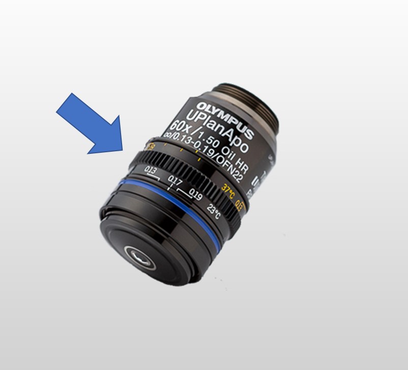
Figure 2. Correction collar on an objective with the indicator for cover glass thickness.
To learn more about motorized lens systems for deep imaging, be sure to read our blog post: How to Improve Deep Imaging in Multiphoton Microscopy.
4. Use a dedicated focusing module to keep samples in focus.
But what if Z-drift occurs?
Even doing all the above, your image might not always be in focus. After all, many factors can cause Z-drift. It can be caused by a proactive and intentional change like adding regents, as well as physical vibration due to someone walking in the room or an environmental temperature change.
And while most motorized microscopes contain an image contrast-based autofocus feature, they have their limitations:
- Limited range of adjustment capability
- Excitation light for contrast-based focusing causes phototoxicity
One way to help ensure focus during live cell experiments is to use a dedicated focusing module like our TruFocus near-infrared (NIR) laser-based Z-drift compensator. NIR light can also help minimize the phototoxicity and crosstalk on the imaging wavelength during prolonged observation.
While the module’s one-shot autofocus (AF) mode prevents unnecessary illumination, continuous AF mode can be used for live observation or continuous image acquisition of fast phenomena.
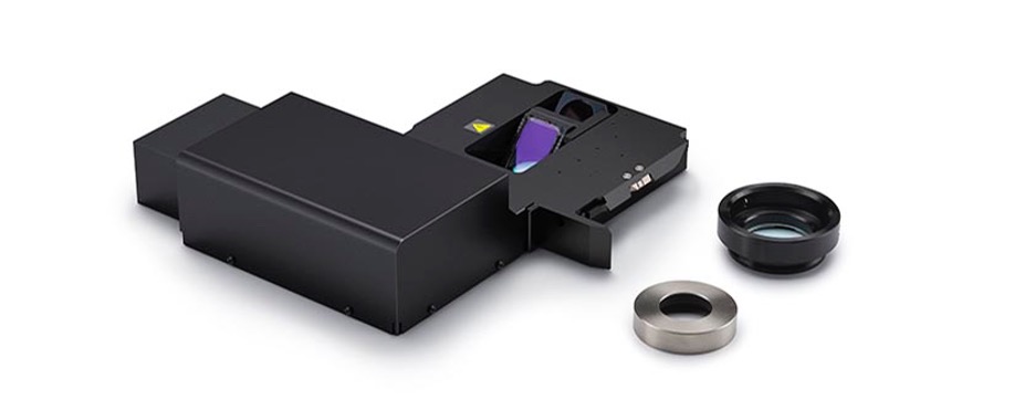
Figure 3. Keep your samples in sharp focus using the Olympus TruFocus Z-drift compensator.
You can see how Z-drift compensation works for long-term imaging by watching the brief video below:
Stay Productive by Running Efficient Live Cell Experiments
There’s always work to be done—so we hope these tips help you stay productive, achieve longer live cell imaging, and continue your science. And if you have any questions about how to automate your lab, don’t hesitate to reach out!
Related Content
How to Improve Deep Imaging in Multiphoton Microscopy
6 Tips for Fluorescence Live Cell Imaging
