
Sharing the stories of our Global Image of the Year (IOTY) contest winners has become an annual tradition, starting with the three regional winners and then culminating with the global winner. So far, for the 2021 competition, we’ve profiled Ivan Radin, the winner for the Americas.
For Europe, the Middle East, and Africa (EMEA), the winning image belongs to Vasilis Kokkoris, a mycologist/ecologist from the Netherlands. Read on to learn more about the image and the research behind it from the microphotographer himself.
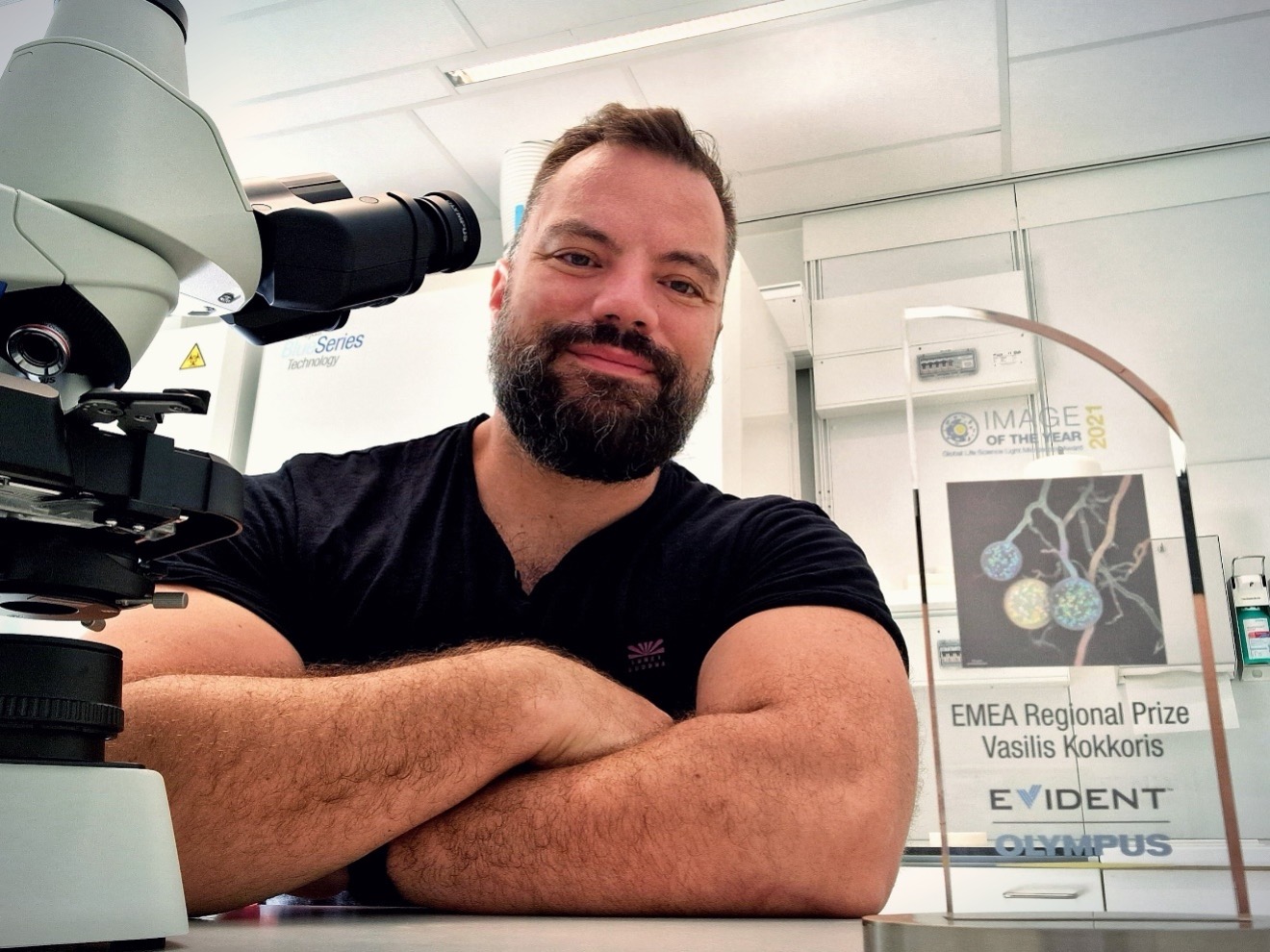
Vasilis Kokkoris, the EMEA regional winner of our Global IOTY 2021 contest with a CX23 microscope
Q: What does your winning image show?
Vasilis: The image depicts three spores (circular formations) of a particular fungal group known as arbuscular mycorrhizal (AM) fungi. Every colourful bright spot within the spores corresponds to a single nucleus. The more abstract lines behind the spores depicts the filamentous body of the fungus—these structures are known as hyphae and they are similar to open pipes where nutrients, water, and organelles travel with impressive speed at long distances.
Q: What do you find most interesting about the subject in your image?
Vasilis: What is unique about these spores is that in contrast to most cells that usually have one or two nuclei, these cells (aka spores) carry hundreds of nuclei and are one of the most multinucleate cells on our planet. I find fascinating the fact that we can view the massive amount of DNA, organized in the nuclei, that each spore carries.
Q: Can you tell us a bit about your current position and the work you do?
Vasilis: I am currently an assistant professor at the Vrije Universiteit (VU) Amsterdam, part of Amsterdam Institute for Life and Environment (A-LIFE). Microscopy is a crucial part of my research. I use it for visualization and quantification of fungal traits and to unlock the genetic secrets of fungi.
My research focuses on better understanding the evolutionary significance of these soil fungi (AM fungi) and their unique nuclear organization and dynamics. I am interested in applying the acquired knowledge towards a more sustainable agriculture and to better understand the challenges that the mycorrhizal symbiosis faces against climate change. For more information, visit my website: www.vasilis-kokkoris.com/.
Q: What sort of imaging tools do you use in your work?
Vasilis: I employ multiple methods to study mycorrhizal fungi. I like to explore unconventional tools that were not destined to be used for fungal research, but that can provide precious information when implemented in my field (mycology and ecology). For example, in recent work (Kokkoris et al., 2019), I used neuron mapping tools to quantify the hyphal growth of mycorrhizal fungi. And in another (Kokkoris Hart, 2019), I used tools that examine root morphology to quantify the presence of mycorrhizal fungi inside the roots. I also use more advanced microscopy (confocal microscopy, scanning electron microscopy, and transiting electron microscopy) to examine the nuclear organization of AM fungi as well as the architecture of their spores (Kokkoris et al., 2020, 2021).
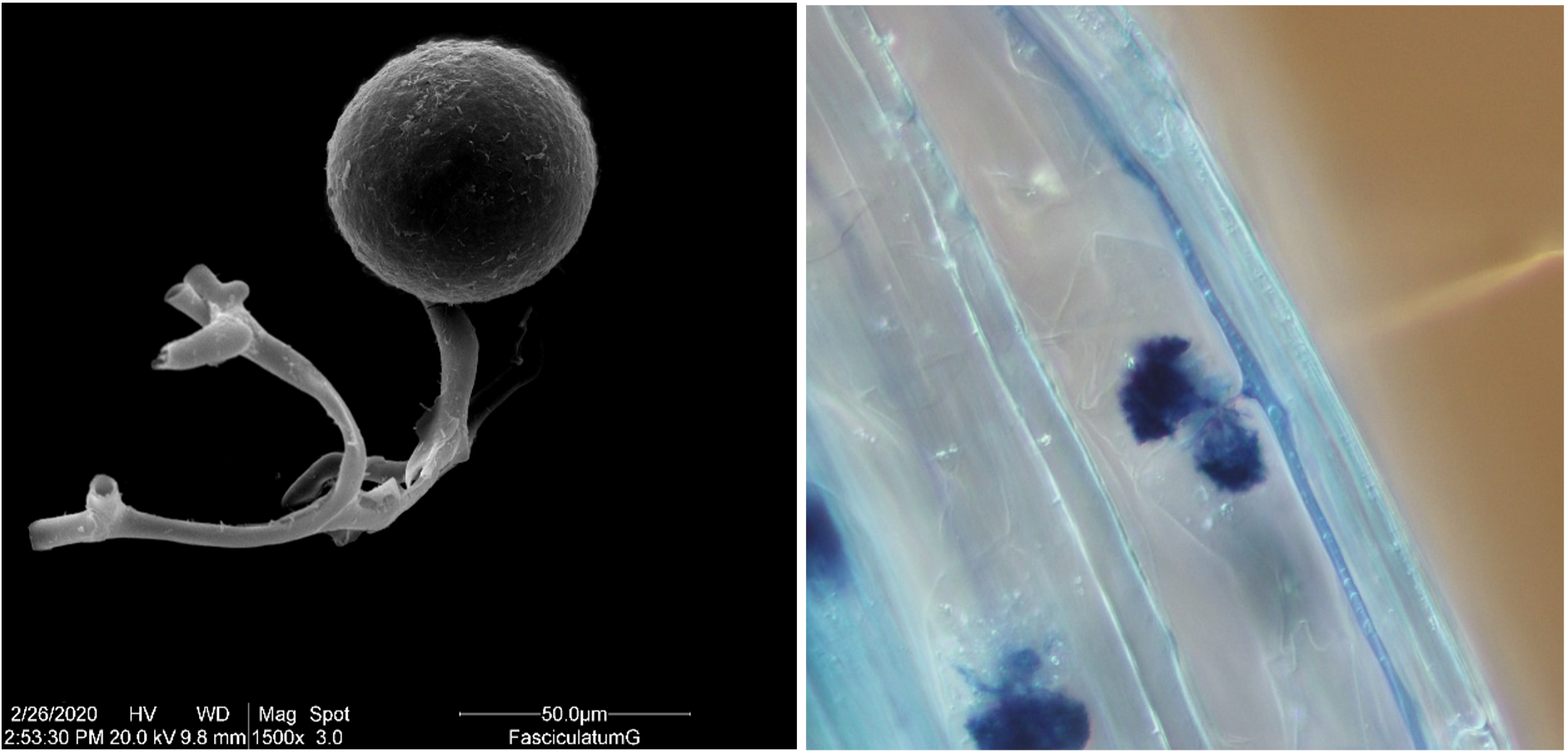
Left: Mycorrhizal spore imaged with scanning electron microscopy. Right: The mycorrhizal fungus (dark blue) colonizes the plant root cells (transparent blue) and forms specialized structures known “arbuscules”. These tree-looking structures are where resources are exchanged between the two partners (plant fungus).
Q: When did you become inspired to use microscopes to create art?
Vasilis: My passion with microscopy started at 12 years old when I received my first microscope as a gift from my parents. It might have been a toy, but it could still magnify specimens up to 40 times. I still remember collecting anything I could possibly find from our garden that would fit between the lens and the stage and examining it under the microscope. That opened my eyes to a fascinating, previously invisible world, and established my passion for biology.
Microphotography has been a huge part of my academic journey. I have been taking photographs through a microscope for many years, trying to use those photographs to extract useful data and scientific conclusions. At the same time, I always devote time to take images which, besides being scientifically useful, also hold artistic merit.
Q: Is there anything else you would like to share?
Vasilis: If you are interested in learning more about the fungal networks and their function and importance, I urge you to visit https://spun.earth/, the site of the Society for the Protection of Underground Networks (SPUN), a group with which I am an associate and core collaborator. SPUN is a nonprofit, science-based initiative founded to map mycorrhizal fungal networks and to advocate for their protection.
References
IOTY 2022 Submissions
If you’re interested in participating in the Global Image of the Year contest for 2022, head to our IOTY page to submit your entry.
Related Content
Old, New, Borrowed, Blue—Our Most Popular Microscope Images for July 2022
Guidelines for Citing Microscopy in Imaging Publications
Aesthetics of Autofluorescence—Meet the IOTY 2020 Global Winner
