
November was a busy (and beautiful) month for microscopy images. Make sure you check out our Olympus Life Science Instagram account to see all the beautiful images we share. Here are our 5 favorite images for the month of November:
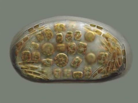
How many different organisms can you spot in this capture? 30? 40? One thing we forgot to mention—these diatom cells are cohabiting in a single drop of water. Talk about a crowded home!
Image courtesy of Dr. Victor Chepurnov.
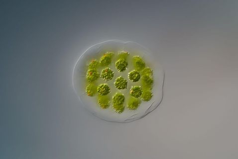
Did you know? Eudorina is a type of green algae that is found in spherical colonies. These colonies typically consist of 16, 32, or 64 individual cells that form groups and move as a whole as the flagellated individual cells work together.
Image courtesy of Håkan Kvarnström.
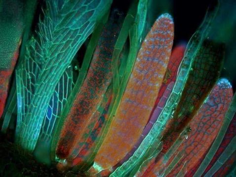
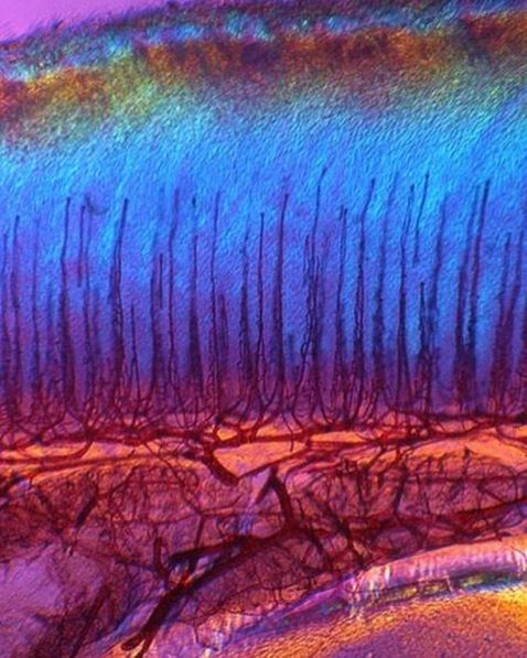
Fluorescence microscopy was used to capture the transverse section of the fertile layer of Mnium hornum (horn calcareous moss).
Image courtesy of Magdalena Turzańska.
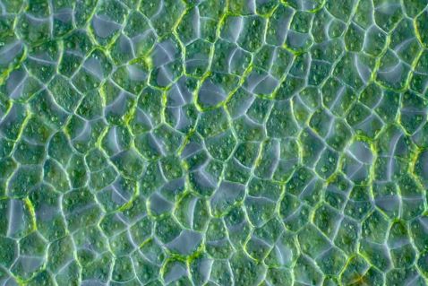
"Lettuce" take a closer look at this algae specimen! Ulva lactuca is a flat, green algae commonly known as sea lettuce that is formed by irregularly arranged layers of cells in a cross-section pattern.
Image courtesy of Linden Gledhill.
Any guesses what animal this tongue belongs to? Here’s a hint: this farm animal was the first egg-laying animal to have its genome sequenced.
Need another hint? This animal descended from the dinosaurs.
Still not sure? It was a hen!
Image courtesy of Dr. Steve Lowry.
Bonus video: This video surpassed all other posts from our account this month!
Related Videos
Instagram user @nomadic_nostoc captured this amazing brightfield video at 1000x magnification using an Olympus CX43 microscope with a new X Line objective. He describes it as follows:
This tardigrade (Hybsius) is hungry and just found the right snack. Hybsius is a vegetarian tardi. It pokes the membrane of the algae (Chlorella) with its stillet organ and slurps the contents right out of the cell. What a cold-blooded murderer... but look at its cute paws! And look at its cute round mouth! Aren’t tardigrades cute?
To see more images like these, be sure to follow us on Instagram at @olympuslifescience!
Interested in sharing your own images?
Visit our image submission site or enter them into our 2019 Global Image of the Year contest.
Related Content
2018 Image of the Year Award Spotlight: 3rd Prize Winner Johann Swanepoel
How to Acquire Microscopy Images that Mimic Human Eye Color Perception
How to Choose the Right Microscope Objective: 10 Questions to Ask
