Distinguishing Brain and Tumor Blood Vessels in Deep Tissues Using Multiphoton Microscopy
Conventional two-photon excitation (2PE) microscopy typically uses laser light in the NIR-I region (700–950 nm) as the excitation light source, limiting the imaging depth to around 500 µm. Compared to NIR-I light, NIR-II light (1000–1700 nm) shows more reduced light attenuation in biological tissues. NIR-II light can also penetrate much deeper to excite the fluorophores during in vivo tissue imaging. In addition, the autofluorescence induced by NIR-II light is much lower than that of NIR-I light, helping to improve the signal-to-background ratio of 2PE images.
Olympus’ FVMPE-RS multiphoton excitation microscope is equipped with an InSight DS pulsed IR laser system to enable multiphoton imaging with excitation from 680–1300 nm. Here are two in vivo deep imaging applications that benefit from using a multiphoton excitation microscope with NIR-II light.
1. NIR-II Excitable Conjugated Polymer Dots for Deep In Vivo Two-Photon Brain Imaging Through an Intact Skull[1]
2PE microscopy through an intact mouse skull is challenging due to strong scattering induced by skull bone. To overcome this challenge, we developed NIR-II light excitable single-chain conjugated polymer dots (CPdots) with bright fluorescence in the NIR-I region (peak at ≈725 nm and quantum yield of 20.6 ± 1.0%) for deep in vivo 2PE imaging of an intact mouse brain. Using Olympus FVMPE-RS equipped with ultrafast tunable laser system, we performed 2PE imaging of the mouse brain during excitation at 800, 1040, and 1200 nm. This system allowed us to tune laser wavelength freely without complicated adjustment work. The 1200 nm excitation led to the largest imaging depth and highest signal-to-background ratio. Moreover, we obtained a 3D reconstruction of the brain blood vessel network with a large vertical depth of 400 µm through the intact skull.
|
|
|
|
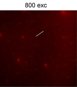
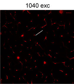
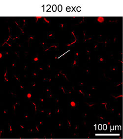
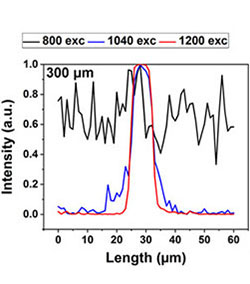
Figure 1. 2PE images of the brain vasculature in a mouse injected with CPdots acquired in the same cortical column (at a depth of 300 µm) during 800, 1040, and 1200 nm fs-laser excitation. Emission was collected within 660–750 nm. Each frame was acquired in 3.22 s. All the images share the same scale bar: 100 µm. The 2PE line intensity profiles across the blood vessels from each frame were plotted. Copyright 2019, Wiley-VCH.
|
|
|
|
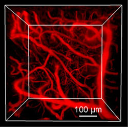
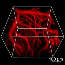
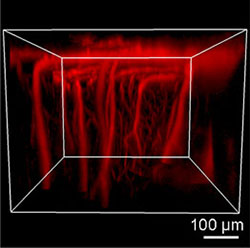
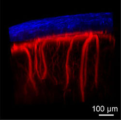
Figure 2. 3D reconstructed 2PE images of the brain blood vessels through an intact skull. Excitation for second harmonic generation (SHG, blue color): 950 nm; Excitation for 2PE: 1200 nm. Emissions were collected within 455–500 nm for SHG and 660–750 nm for blood vessels. Each frame was acquired in 3.22 s. Copyright 2019, Wiley-VCH.
Related Videos |
Video 1. 3D reconstructed 2PE image of the brain blood vessels labeled by CPdots. Excitation: 1200 nm. Emission: 660–750 nm. Copyright 2019, Wiley-VCH.
2. NIR-II-Excited Intravital Two-Photon Microscopy Distinguishes Deep Cerebral and Tumor Vasculatures[2]
Intravital fluorescence imaging of vasculature morphology and dynamics in the brain and tumors with a large penetration depth and high signal-to-background ratio (SBR) is ideal for the study and theranostics of vascular-related diseases and cancers. A highly bright fluorophore with aggregation-induced NIR emission is designed for NIR-II light excited intravital 2PE microscopy of brains and tumors. Due to the deep penetration capability of NIR-II light and high brightness of the fluorophore, we could image the vasculatures in deep tissues (brain and tumors).
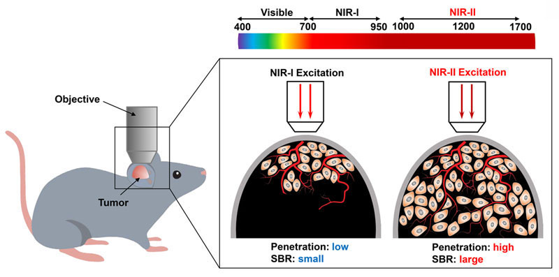
Scheme 1. Schematic illustration of in vivo two-photon fluorescence imaging of a tumor under NIR-I and NIR-II excitation. Copyright 2019, Wiley-VCH.
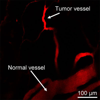
| Figure 3. 2PE image of a tumor and normal blood vessels labeled by our fluorophore. The tumor blood vessel shows enhanced 2PE compared to the normal vessel. Excitation: 1200 nm; Emission: 660–750 nm. Copyright 2019, Wiley-VCH. |
|---|
|
|
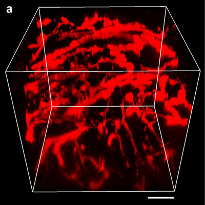
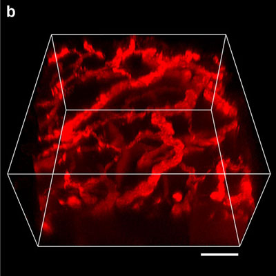
Figure 4. 3D reconstructed 2PE images of tumor vasculature networks in the same tumor under NIR-II (a) and NIR-I (b) excitation. Excitations: 1200 nm for NIR-II and 920 nm for NIR-I. Scale bars: 100 μm. Imaging depth (through skin): 500 μm for NIR-II excitation and 300 μm for NIR-I excitation. Copyright 2019, Wiley-VCH.
References:
[1] NIR‐II Excitable Conjugated Polymer Dots with Bright NIR‐I Emission for Deep In Vivo Two‐Photon Brain Imaging Through Intact Skull
[2] NIR-II-Excited Intravital Two-Photon Microscopy Distinguishes Deep Cerebral and Tumor Vasculatures with Ultrabright NIR-I AIE Luminogen
Imaging equipment
Microscope: FVMPE-RS system
Objective: 25X water immersion objective (XLPLN25XWMP2)
Comment by Dr. Shaowei Wang
| The Olympus FVMPE-RS multiphoton microscope is a powerful tool for intravital imaging. The InSight DS pulsed IR laser with tunable wavelengths from 680–1300 nm is useful for various in vivo two-photon fluorescence imaging applications. Due to the optimized optical coating and excellent objective with a high transmittance in the NIR-II region, we could perform NIR-II excited intravital two-photon microscopy with our ultrabright fluorophores. The imaging depth and signal-to-background ratio improved under NIR-II excitation. |

Acknowledgments
This application note was prepared with the help of the following researcher:
Dr. Shaowei Wang, Department of Chemical and Biomolecular Engineering, National University of Singapore (Homepage)
How FVMPE-RS facilitated our experiment
| Efficient Laser Transmission at NIR-IISilver-coated scanner mirrors help deliver more laser power to your sample for brighter images. Increased reflectance in the near-infrared range compared to conventional aluminum-coated mirrors is especially advantageous for deep in vivo experiments. |
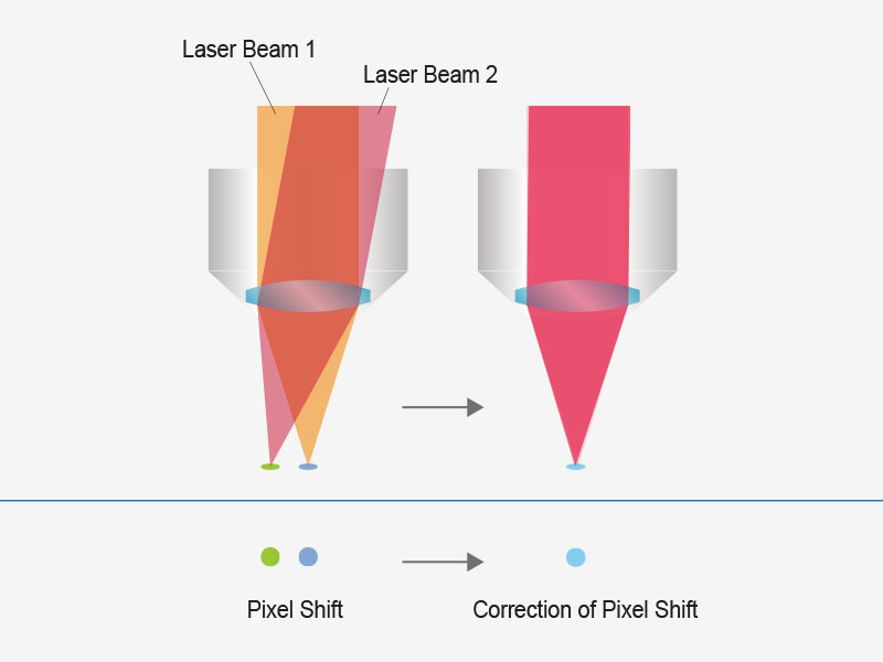
| User-Friendly Imaging with Automated Laser AlignmentLaser drift caused by wavelength tuning can cause a misalignment of excitation beam that reduces multiphoton image quality in typical systems. |

Products related to this application
Sorry, this page is not
available in your country.