Monitoring the Effect of Medium Components on the Growth and Differentiation of Human iPS Cell-Derived Liver Bud Organoids Using the Olympus Provi™ CM20 Incubation Monitoring System
Introduction
Organoids are used in developmental biology research to understand organogenesis and in drug discovery research to elucidate the pathogenic mechanism of diseases and find an effective treatment. Optimizing culture conditions such as growth factors and small molecule compounds is essential for successfully culturing organoids. Currently, the optimal culture conditions are achieved through trial and error, and it is important to carefully observe the changes in the behavior and morphology of the cells cultured under each condition. In this application note, we show how the Olympus Provi™ CM20 delivers qualitative results that are essential for establishing the optimal culture conditions for liver bud organoids.
Experimental Outline
The creation of liver bud organoids derived from human iPS cells has shown potential value in a variety of settings (Koido M et al., Nature Medicine 2020 (PMID: 32895570), Takebe T et al., Nature 2013 (PMID: 23823721)). For this experiment, liver bud organoids were differentiated from human iPS cells in a three-dimensional culture system. Five different conditions in which medium components were modified to alter later stages of liver bud differentiation (named here A, B, C, D, and E) were tested. The differentiation and morphology of the organoids were analyzed to establish the effect of the different compounds and find the optimal culture condition. The CM20 incubation monitoring system was used to acquire information on the 3D cultured organoids placed in a 24-well plate for a period of about 2 weeks (Figure 1).
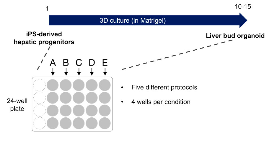
Figure 1. Overview of the experimental conditions
Results
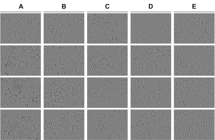
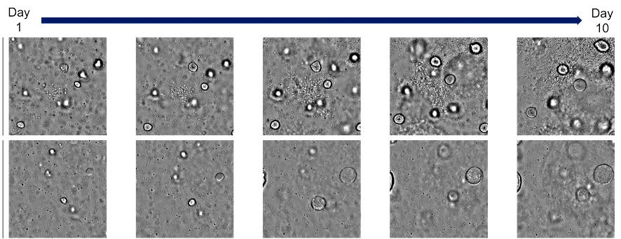
Under the already established condition A, it was observed that spherical organoids were formed and self-organized structures gradually developed. In conditions B and C, it was observed that morphologically similar organoids to those in A were formed. On the other hand, applying conditions D and E led to an increase in the diameter and quantity of the organoids (Figure 2). The temporal data obtained with the CM20 system also revealed that the luminal dilation observed under conditions D and E occurred approximately 4–5 days after the compound addition (Figure 3).
Figure 2. Observation of human liver bud organoids grown in a multiwell plate and exposed to five different small compounds that affected their differentiation and growth |
Figure 3. Morphological changes in human liver bud organoids in response to compound addition |
Conclusion
In conclusion, it was found that by using the CM20 monitoring system, the state of three-dimensional organoids cultured in multiwell plates can be monitored over time, and comparative observation between conditions is possible.
Comments from Drs. Takebe and Yoneyama


Dr. Takanori Takebe (left), Dr. Yosuke Yoneyama (right) Tokyo Medical and Dental University Institute of Research.
Although it is important to analyze the expression of molecular markers in evaluating the efficiency of organoid differentiation, the morphological changes in organoids can also be useful indicators of their quality and future results. With the CM20 system, the state of organoids is constantly monitored, so we don’t miss the timing of when morphological changes occur.
Products Related to This Application
Sorry, this page is not
available in your country.