Monitoring the Whole Process of Human iPS Cell-Derived Liver Bud Organoid Differentiation Using the CM20 Incubation Monitoring System
Introduction
Recently, rapid progress has been made in in vitro establishment of miniature tissues/organs, called organoids, from human pluripotent stem cells including induced pluripotent stem (iPS) cells. Human organoids provide unique opportunities for a wide range of fields, from basic developmental biology to disease modeling and drug development.
Experimental background
In general, organoids are formed through self-organization, in which composing cells are spatially arranged in appropriate locations to determine their differentiation fates into specific cell types, eventually leading to the expression of organ functions. The process of deriving undifferentiated human iPS cells to the organoids of a target organ requires a long period of time, at least a month or more. The long duration is due to the existence of multiple stages of intermediate differentiated cells corresponding to developmental stages. In many cases, these processes also include the steps of embedding the cells into an extracellular matrix containing basement membrane components, which is essential for polarization of cells and subsequent maintenance of three-dimensional structures. In other words, successfully and reproducibly generating organoids of a certain quality from human iPS cells inevitably requires careful examination of both two- and three-dimensional culture conditions. Specifically, in addition to the quality control of iPS cells, it is important to seamlessly monitor the differentiation processes over a long period of time, including data gathering on three-dimensional behaviors and morphogenesis of cell populations.
In this application note, we attempted to observe the differentiation process using the Olympus Provi CM20 incubation monitoring system. We used our recently published method for generating human liver bud organoids1,2 as a model example.
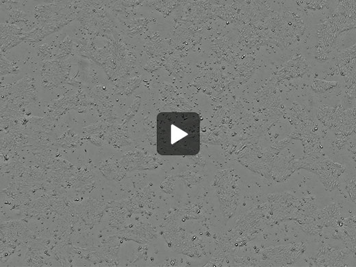
Observation of Morphological Changes during the Formation of the Posterior Foregut
Using the CM20 system, we tested whether it is possible to observe three-dimensional morphological changes in the differentiation process of undifferentiated human iPS cells into a two-dimensional sheet-like definitive endoderm and the subsequent emergence of posterior foregut spheroids.
|
Results
Using the CM20 system, we were able to observe not only the process of human iPS cells culture maintenance, but also their differentiation into definitive endoderm and the subsequent emergence of posterior foregut spheroids. These posterior foregut spheroids were generated through the cell-autonomous formation of three-dimensional cell aggregates derived from the two-dimensional cell sheet, demonstrating that the process of such morphological changes can be tracked with CM20 system. Since organoids of other endodermal organs, such as the stomach and intestine, are also derived from primitive gut spheroids originated from the definitive endoderm, we propose that the CM20 system can be broadly applied to monitor the differentiation process of such organoids.
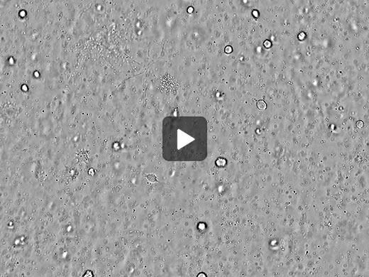
Observation of the Culture Process of Liver Bud Organoids in Matrigel
Using the CM20 system*1, we tested whether it is possible to observe the culture process of liver bud organoids that differentiate and grow in Matrigel in a three-dimensional manner.
|
Results
Using the CM20 system, we were able to follow the differentiation process of liver bud organoids in a three-dimensional culture using Matrigel drops in a 24-well plate over about 2 weeks.
Conclusion
Using the automated monitoring features of the CM20 system’s software, it is possible to track the differentiation process that causes morphological changes as well as the growth process of three-dimensional organoids in extracellular matrix gel. Our experiment showed that the CM20 system can monitor the long-term culture process—from the maintenance and management of human iPS cells to their differentiation into organoids—and can contribute to the optimization and quality control of cell cultures.
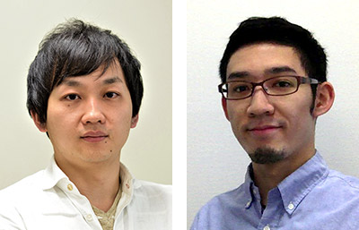
Comments from Dr. Takebe and Dr. Yoneyama
Dr. Takanori Takebe (left) | We were impressed that the CM20 monitoring system enabled us to track spheroid formation and organoids cultured in extracellular matrix gel with fine contrast. Many researchers must be curious how the differentiation and formation process of organoids, which requires long-term culture, takes place in the incubator. We believe that this machine can be used for research in a wide range of cell biology-related fields. |
References
- Koido M et al., Nature Medicine 2020 (PMID: 32895570), Takebe T et al., Nature 2013 (PMID: 23823721), Takebe T et al., Cell reports 2017 (PMID: 29212014)
- Tadahiro Shinozawa, Masaki Kimura, Yuqi Cai, Norikazu Saiki, Yosuke Yoneyama, Rie Ouchi, Hiroyuki Koike, Mari Maezawa, et al., “High-Fidelity Drug-Induced Liver Injury Screen Using Human Pluripotent Stem Cell-Derived Organoids.” 2021 Feb;160(3):831-846.e10. doi: 10.1053/j.gastro.2020.10.002. Epub 2020 Oct 8. PMID: 33039464 PMCID: PMC7878295 (available on 2022-02-01) DOI: 10.1053/j.gastro.2020.10.002
*1 Using features added to the version 1.2.5 or later of the Olympus Provi CM20 system. Download the update or read the software release notes for details.
Products related to this application
Sorry, this page is not
available in your country.