Evident LogoOlympus Logo
Application Notes

Single-Molecule Fluorescence Imaging on the Cell Membrane


Single-Molecule Fluorescence Imaging on the Cell Membrane Using a Super High Numerical Aperture (NA) Objective Lens

Introduction

Recent advances in cell preparation and microscope optical systems have enabled imaging of single biomolecules in a live cell. Molecular dynamics, such as the binding of a physiologically active ligand to a cell, dimerization of signal molecules, and the formation of a molecular complex, can be visualized at the single molecule level in live a cell using objective lenses with a super high numerical aperture. In this study, researchers used an Olympus super high NA objective lens for fluorescence imaging of intermolecular interactions in ion channels on the cell membrane at the single molecule level.

Super high NA objective lens TIRF application

Fluorescent-protein (FP) tagged ion channel subunits are expressed in Xenopus oocytes and observed at the single molecule level by TIRF microscopy (Figure 1, left). Stochastic bleaching events of individual FPs can be observed as ‘bleaching steps’ (Figure 1, right). The number of subunits in a single ion channel complex can be determined by counting the bleaching steps from individual fluorescent spots.

Figure 1. A schematic overview of the subunit counting by single molecule photobleaching. The photobleaching steps are represented by the green arrows.

Figure 2. Images of the fluorescent proteins Kv4.2-mCherry (left) and mEGFP-DPP10 (middle) expressed in Xenopus oocyte at the single molecule level under TIRF microscopy.
Figure 2. Images of the fluorescent proteins Kv4.2-mCherry (left) and mEGFP-DPP10 (middle) expressed in Xenopus oocyte at the single molecule level under TIRF microscopy. 

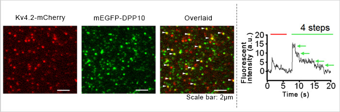
Images of the fluorescent proteins Kv4.2-mCherry Figure 2, left) and mEGFP-DPP10 (Figure 2, middle) expressed in a Xenopus oocyte were observed at the single molecule level using TIRF microscopy and super high NA objective lenses.  Each red spot represents a single Kv4.2 channel (tetramer).  Some of the green spots overlap with the red spots (white arrowheads in Figure 2, right) indicating that Kv4.2 and DPP10 form a complex.  By counting bleaching events of mEGFP from a single fluorescent spot, the number of subunits in the complex can be counted. 1  In the Figure 2 graph, four bleaching events (green arrows) were observed from a Kv4.2-mCherry/mEGFP-DPP10 spot, suggesting four DPP10 subunits were included in the complex.
1 Ulbrich, Maximilian H., and Ehud Y. Isacoff. “Subunit counting in membrane-bound proteins.” Nature methods 4, no. 4 (2007): 319–321


Movie of Kv4.2-mCherry and mEGFP-DPP10

Imaging System;
Microscope: Research Inverted Microscope IX71
Objective:  Apo 100XOHR (100X, N.A.1.65)
Ex: 488nm (Solid laser, Spectra-Physics) , 588nm (Solid laser, Coherent)
CCD camera: iXon3 EMCCD camera (Andor)
Coverslips: High refractive index coverslip (n = 1.78)

Image data courtesy of;
Masahiro Kitazawa, Ph.D., Yoshihiro Kubo, M.D.,Ph.D., Koichi Nakajo*, Ph.D.
Division of Biophysics and Neurobiology, Department of Molecular Physiology, National Institute for Physiological Sciences
*Present address:  Department of Physiology, Osaka Medical College

Reference;
J Biol Chem. 2015 Sep 11; 290(37):22724-33. doi: 10.1074/jbc.M115.646794.
J Biol Chem. 2014 Jun 20;289(25):17597-609. doi: 10.1074/jbc.M114.563452.
Proc Natl Acad Sci U S A. 2010 Nov 2;107(44):18862-7. doi: 10.1073/pnas.1010354107.

Conclusion

A high numerical aperture objective lens designed only for evanescent illumination can produce remarkably high contrast images even with weak fluorescent light because of the efficient formation of an evanescent wave field with a shallow penetration depth. While this type of observation requires the quantitative measurement of minute changes in fluorescence intensity due to fluorescence loss at the single molecule level, observation using a super high NA objective lens with special immersion oil and coverslips facilitates images of intermolecular interactions in ion channels in Xenopus oocyte membranes. Since these images have a high signal-to-noise ratio, changes in fluorescence intensity can be quantitatively measured.

Products related to this application

TIRF Imaging Microscope System

IXplore TIRF

For membrane dynamics, single-molecule detection, and colocalization experiments, the IXplore TIRF system enables sensitive simultaneous multicolor TIRF (total internal reflection fluorescence) imaging for up to four colors. Olympus’ cellTIRF system provides stable motorized individual laser-angle control, providing equal evanescent wave penetration for high-contrast, low noise images. Our TIRF objectives feature high SNR, high NA, and correction collars to adjust for cover glass thicknesses and temperature.

  • Exact colocalization of up to four markers thanks to individual penetration depth control
  • Take advantage of Olympus’ TIRF objective with the world's highest NA of 1.7*
  • Intuitive setup of complex experiments with the Graphical Experiment Manager (GEM), cellFRAP, and U-RTCE
* As of July 25, 2017. According to Olympus research.
Apochromat Objectives for Super Resolution/TIRF Imaging

APON-TIRF/UAPON-TIRF/UPLAPO-HR

Offering our highest numerical aperture values, these apochromat objectives are optimized for high-contrast TIRF and super resolution imaging. Achieve wide flatness with the UPLAPO-HR objectives’ high NA, enabling  real-time super resolution imaging of live cells and micro-organelles.

  • High NA to create an evanescent wave field for high contrast TIRF images or super resolution
  • HR series are the world first* Plan Apochromat objectives with NA1.5 which achieve wide flatness

* As of November 2018. According to Olympus research.

was successfully added to your bookmarks

View BookmarksClose

Maximum Compare Limit of 5 Items

Please adjust your selection to be no more than 5 items to compare at once

Sorry, this page is not
available in your country.

Sorry, this page is not available in your country