Evident LogoOlympus Logo

Ask the Experts

Light Sheet Microscopy for Deeper Insight into Life

Light Sheet Microscopy for Deeper Insight into Life

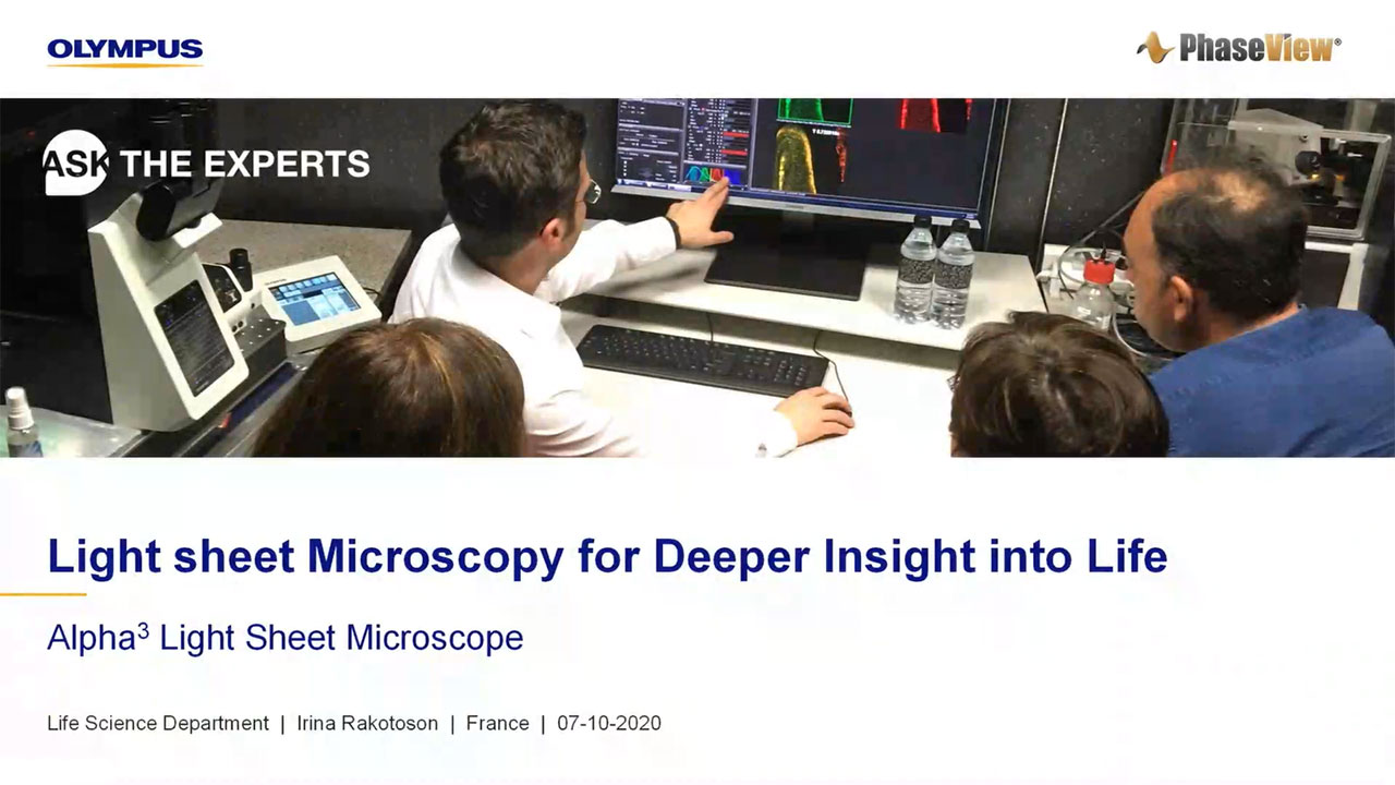
This webinar introduces you to light sheet microscopy. The Alpha3 light sheet microscope combines very thin optical sectioning and high-quality Olympus optics for high-resolution 3D imaging of both live and fixed biological samples. The Alpha3 microscopy system integrates cutting-edge technologies that offer unprecedented image quality at enhanced subcellular resolution. In this webinar, you will discover a highly versatile and modular light sheet system for either routine or high-end research, from macro to micro imaging of whole organs to very small specimens.

Presenter:

Irina Rakotoson, Life Science Product Manager

FAQ

Webinar FAQs | Light sheet microscopy for deeper insight into life

What’s the maximum resolution of the Alpha3 system in the x-y plane and along the z axis?

The maximum resolution in the x-y plane is given by the physical length of pixels on the camera divided by the objective’s magnification. For example, at 40X the maximum resolution is 6.5 µm/40 = 0.16 µm.

In light sheet microscopy, the maximum resolution along the z axis is limited by the width of the illuminating light sheet—the thinnest light sheet possible in the Alpha3 system is 2 µm. For magnifications over 10X, every 1 micron of the sample can be scanned. Nyquist sampling then limits the maximum resolution in the z plane to twice this, which is 2 µm.

What types of lasers are used in light sheet microscopy, and what is their average power rating?

We can integrate from 4 to 6 laser lines into the combiners. These are mostly laser diodes, with the exception of the 561 line, which is a DPS that requires a modulator to set up the power. The average power rating is usually between 50 and 100 mW.

Can the Alpha3 system have a dual camera setup?

Most configurations are flexible enough with one camera, although two camera setups are also possible.

Can the Alpha3 system do multicolor imaging?

There are two multicolor imaging methods with the Alpha3 system. The first involves scanning the same region of the sample several times and changing the laser and emission filter each time.

The second is ‘flashing mode,’ which uses a rejection filter after the objective that lets the whole broadband spectrum through but cuts out the laser lines. This way we can scan the sample once and flash each laser for each layer. This method is quicker and provides much better colocalization for live samples.

Can IR lasers be integrated with the Alpha3 system for multiphoton light sheet microscopy?

Currently, 730 nm is the longest wavelength of the visible spectrum available, but we are exploring multiphoton applications.

Can conventional deconvolution algorithms be used on data generated by the Alpha3 system?

Yes, deconvolution can be performed on the data generated, and this analysis has previously been performed by Olympus.

Can images taken by the Alpha3 system be exported into an open-source platform for deconvolution?

The software included with the Alpha3 system generates images that can be opened by any open-source software. However, the system used for image processing must be able to load and process large datasets of 300–800 GBs.

Are there special requirements for data handling or image storage in light sheet microscopy?

The Alpha3 system comes with a computer with enough memory to handle large datasets, but we recommend using a sever for long-term storage. It is also best to upload the data to a server as quickly as possible after acquisition.

What is the fastest acquisition speed possible using the optical focus sweeping?

The optical focus sweeping will slow down the acquisition rate by a factor of 4. For example, for an exposure time of 10 ms, you can have a frame time of 40 ms, which will provide 20 frames/second.

How easy is it to sterilize the sample holders and the clamps?

All the parts are made of medical-grade stainless steel and are chemical and corrosion resistant and inert. They can be autoclaved or submerged in pure ethanol to sterilize.

Which camera is included with the Alpha3 system?

The system comes with the ORCA-flash 4.0 digital CMOS camera. It has 6.5 µm pixels and a 2048 × 2048 sensor.

Does the Alpha3 system come with a fast network card for data transfer?

We provide 1, 10, and 100 GHz options for data transfer, but we can also meet custom requests.


Related Products

Light Sheet Microscope

Alpha3

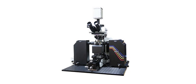
The Alpha3 advanced fluorescence light sheet microscope provides researchers with a flexible, high-performing light sheet solution. Using smart optical focus sweeping, it acquires uniform, artifact-free imaging across the entire field of view. Easily upgrade the Alpha3 system to meet your specific needs by selecting from options such as Smart 3D scanning, XY tiling, or temperature controls, based on your goals.

  • Multidirectional light sheet with real-time focus sweeping for rapid, artifact-free 3D imaging
  • Compatible with a large selection of high-quality Olympus optics
  • Accessories support a variety of specimens and are resistant to organic solvents or aqueous buffers
  • User-friendly QtSPIM software and workstation enables seamless 3D image acquisition at maximum speed

Light Sheet Microscopy for Deeper Insight into Life

This webinar introduces you to light sheet microscopy. The Alpha3 light sheet microscope combines very thin optical sectioning and high-quality Olympus optics for high-resolution 3D imaging of both live and fixed biological samples. The Alpha3 microscopy system integrates cutting-edge technologies that offer unprecedented image quality at enhanced subcellular resolution. In this webinar, you will discover a highly versatile and modular light sheet system for either routine or high-end research, from macro to micro imaging of whole organs to very small specimens.

Large sample mounting procedure

Small sample mounting procedure

Key technologies enable superior image quality

3D image of whole zebrafish brain

3D image of whole zebrafish larva

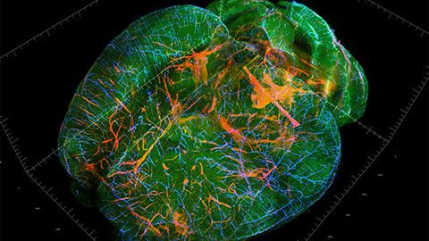
3D image of whole mouse brain

FAQ

Webinar FAQs | Light sheet microscopy for deeper insight into life

What’s the maximum resolution of the Alpha3 system in the x-y plane and along the z axis?

The maximum resolution in the x-y plane is given by the physical length of pixels on the camera divided by the objective’s magnification. For example, at 40X the maximum resolution is 6.5 µm/40 = 0.16 µm.

In light sheet microscopy, the maximum resolution along the z axis is limited by the width of the illuminating light sheet—the thinnest light sheet possible in the Alpha3 system is 2 µm. For magnifications over 10X, every 1 micron of the sample can be scanned. Nyquist sampling then limits the maximum resolution in the z plane to twice this, which is 2 µm.

What types of lasers are used in light sheet microscopy, and what is their average power rating?

We can integrate from 4 to 6 laser lines into the combiners. These are mostly laser diodes, with the exception of the 561 line, which is a DPS that requires a modulator to set up the power. The average power rating is usually between 50 and 100 mW.

Can the Alpha3 system have a dual camera setup?

Most configurations are flexible enough with one camera, although two camera setups are also possible.

Can the Alpha3 system do multicolor imaging?

There are two multicolor imaging methods with the Alpha3 system. The first involves scanning the same region of the sample several times and changing the laser and emission filter each time.

The second is ‘flashing mode,’ which uses a rejection filter after the objective that lets the whole broadband spectrum through but cuts out the laser lines. This way we can scan the sample once and flash each laser for each layer. This method is quicker and provides much better colocalization for live samples.

Can IR lasers be integrated with the Alpha3 system for multiphoton light sheet microscopy?

Currently, 730 nm is the longest wavelength of the visible spectrum available, but we are exploring multiphoton applications.

Can conventional deconvolution algorithms be used on data generated by the Alpha3 system?

Yes, deconvolution can be performed on the data generated, and this analysis has previously been performed by Olympus.

Can images taken by the Alpha3 system be exported into an open-source platform for deconvolution?

The software included with the Alpha3 system generates images that can be opened by any open-source software. However, the system used for image processing must be able to load and process large datasets of 300–800 GBs.

Are there special requirements for data handling or image storage in light sheet microscopy?

The Alpha3 system comes with a computer with enough memory to handle large datasets, but we recommend using a sever for long-term storage. It is also best to upload the data to a server as quickly as possible after acquisition.

What is the fastest acquisition speed possible using the optical focus sweeping?

The optical focus sweeping will slow down the acquisition rate by a factor of 4. For example, for an exposure time of 10 ms, you can have a frame time of 40 ms, which will provide 20 frames/second.

How easy is it to sterilize the sample holders and the clamps?

All the parts are made of medical-grade stainless steel and are chemical and corrosion resistant and inert. They can be autoclaved or submerged in pure ethanol to sterilize.

Which camera is included with the Alpha3 system?

The system comes with the ORCA-flash 4.0 digital CMOS camera. It has 6.5 µm pixels and a 2048 × 2048 sensor.

Does the Alpha3 system come with a fast network card for data transfer?

We provide 1, 10, and 100 GHz options for data transfer, but we can also meet custom requests.


Related Products

Light Sheet Microscope

Alpha3

The Alpha3 advanced fluorescence light sheet microscope provides researchers with a flexible, high-performing light sheet solution. Using smart optical focus sweeping, it acquires uniform, artifact-free imaging across the entire field of view. Easily upgrade the Alpha3 system to meet your specific needs by selecting from options such as Smart 3D scanning, XY tiling, or temperature controls, based on your goals.

  • Multidirectional light sheet with real-time focus sweeping for rapid, artifact-free 3D imaging
  • Compatible with a large selection of high-quality Olympus optics
  • Accessories support a variety of specimens and are resistant to organic solvents or aqueous buffers
  • User-friendly QtSPIM software and workstation enables seamless 3D image acquisition at maximum speed
Experts
Irina Rakotoson
Life Science Product Manager, PhaseView

Hello. I’m Irina Rakotoson and I’m the Life Science Product Manager for PhaseView. I obtained my master's degree in Cell Biology, Physiology and Pathology specialized in aging at Paris Descartes University. Before joining PhaseView as Product Manager, I worked in a Neuroscience research lab (SPPIN), optimizing clearing protocols for the microscopy core facility.

Light Sheet Microscopy for Deeper Insight into LifeApr 18 2024
Sorry, this page is not available in your country
Ask the Expert Sign-up

By clicking subscribe you are agreeing to our privacy policy which can be found here.

Sorry, this page is not
available in your country.

Sorry, this page is not available in your country