Evident LogoOlympus Logo

Ask the Experts

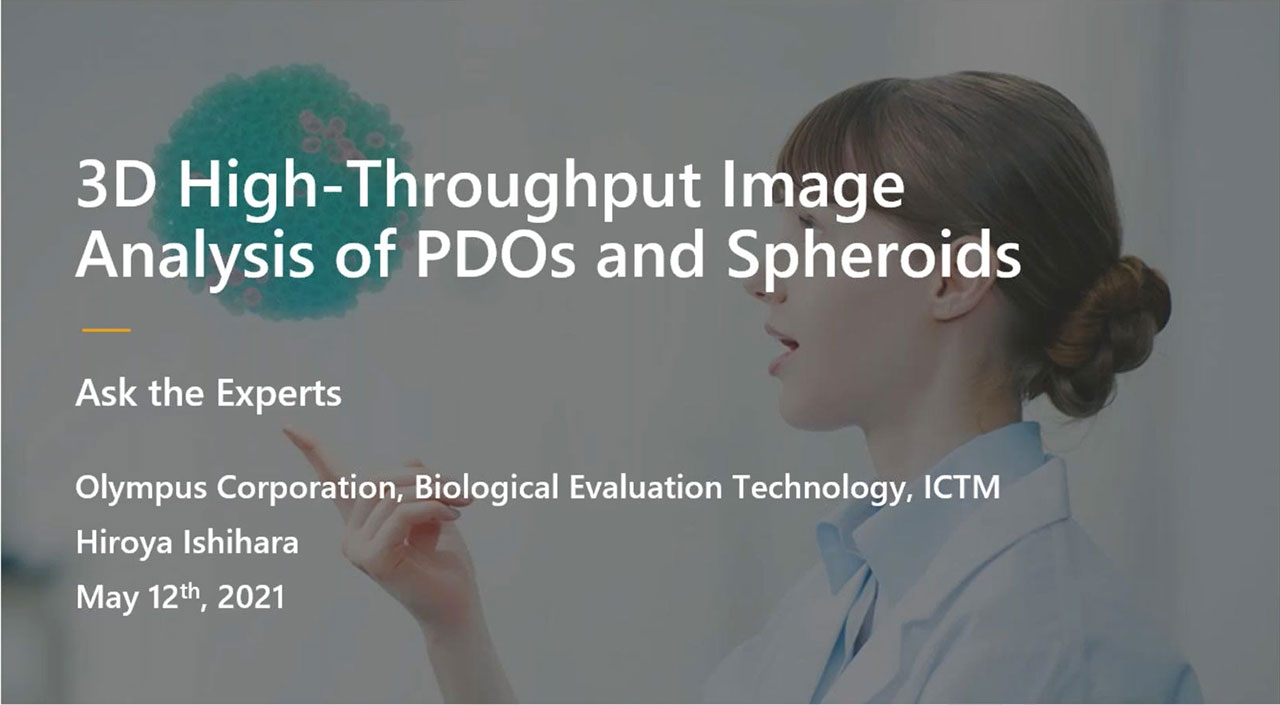
Three-Dimensional High-Throughput Image Analysis of Patient-Derived Organoids and Spheroids

Three-Dimensional High-Throughput Image Analysis of Patient-Derived Organoids and Spheroids

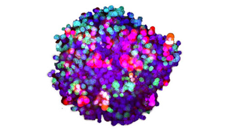
Organoids and spheroids can more faithfully reproduce in vivo conditions than standard 2D cell cultures, and imaging-based analysis can monitor cell-specific responses with high spatial resolution. Therefore, we have been developing imaging-based three-dimensional analysis and drug evaluation methods using patient-derived cancer organoids and spheroids.

In this webinar, we will present some examples of applications within recent trends in organoid research.

Presenters:

Hiroya Ishihara
Applications Scientist Olympus Technologies Asia, Olympus Corporation

FAQ

Webinar FAQs | 3D High-Throughput Image Analysis of PDOs and Spheroids

What sets NoviSight™ software apart from other 3D imaging analysis software?

While other image analysis software may have a graphical user interface and recognize complex 3D structures, NoviSight software is unique for its high-throughput 3D image analysis and intuitive object classification. For instance, NoviSight software is ideal for performing a high-throughput analysis of spheroids and other 3D objects in microplate-based experiments.

Can NoviSight software be used for other 3D imaging applications?

In addition to analysis of organoids and spheroids, NoviSight software can perform measurements based on basic object recognition and classifaction for a range of applications. For example, you can quantify the region expressing a specific gene in 3D organs. However, NoviSight software does not have a module for recognizing filament structures, such as dendrites, so it cannot be used to analyze these objects.

Can NoviSight software be used to analyze 2D objects?

Yes, of course. You can measure 2D objects using general object recognition and classification. If you plan to do only 2D analysis, cellSens™ software may be more suitable because it is optimized for 2D analysis. However, if you want to analyze both 2D and 3D objects, NoviSight software is the ideal choice.

Can NoviSight software be used with widefield microscopy?

3D data acquired with the widefield systems is unsuitable for analysis, as images do not have a high enough resolution. We recommend you use cellSens software to employ the deconvolution process.

Does optical clearing cause tissue shrinkage?

Sometimes the sample shrinks or expands when using a transparent clearing reagent. However, in our experience, the SCALEVIEW-S4 tissue clearing reagent is suitable for organoids and spheroids, and there is no expansion or shrinking.

How much time does it take to count the number of cells in a spheroid?

It takes 10 minutes or less to count the number of cells in a spheroid.

Does NoviSight software include deep-learning technology?

Our TruAI™ deep-learning technology is available in both cellSens software and the scanR high-content screening station but is not available in NoviSight software.

Is a free trial available for NoviSight software?

To try the software for free, contact your local representative to receive a trial version.


Related Products

3D Cell Analysis Software

NoviSight

NoviSight 3D cell analysis software provides statistical data for spheroids and 3D objects in microplate-based experiments. Use it to quantify cell activity in 3D, easily capture rare cell events, obtain accurate cell counts, and improve detection sensitivity. NoviSight software works with a range of imaging techniques, including point-scan confocal imaging, two-photon imaging, spinning disk confocal imaging, and super resolution live cell imaging.

  • Fast 3D image recognition from whole structures to subcellular features
  • Accurate statistical analysis
  • Equipped with a variety of ready-to-use default assays or easily design your own

Three-Dimensional High-Throughput Image Analysis of Patient-Derived Organoids and Spheroids

Organoids and spheroids can more faithfully reproduce in vivo conditions than standard 2D cell cultures, and imaging-based analysis can monitor cell-specific responses with high spatial resolution. Therefore, we have been developing imaging-based three-dimensional analysis and drug evaluation methods using patient-derived cancer organoids and spheroids.

In this webinar, we will present some examples of applications within recent trends in organoid research.

FAQ

Webinar FAQs | 3D High-Throughput Image Analysis of PDOs and Spheroids

What sets NoviSight™ software apart from other 3D imaging analysis software?

While other image analysis software may have a graphical user interface and recognize complex 3D structures, NoviSight software is unique for its high-throughput 3D image analysis and intuitive object classification. For instance, NoviSight software is ideal for performing a high-throughput analysis of spheroids and other 3D objects in microplate-based experiments.

Can NoviSight software be used for other 3D imaging applications?

In addition to analysis of organoids and spheroids, NoviSight software can perform measurements based on basic object recognition and classifaction for a range of applications. For example, you can quantify the region expressing a specific gene in 3D organs. However, NoviSight software does not have a module for recognizing filament structures, such as dendrites, so it cannot be used to analyze these objects.

Can NoviSight software be used to analyze 2D objects?

Yes, of course. You can measure 2D objects using general object recognition and classification. If you plan to do only 2D analysis, cellSens™ software may be more suitable because it is optimized for 2D analysis. However, if you want to analyze both 2D and 3D objects, NoviSight software is the ideal choice.

Can NoviSight software be used with widefield microscopy?

3D data acquired with the widefield systems is unsuitable for analysis, as images do not have a high enough resolution. We recommend you use cellSens software to employ the deconvolution process.

Does optical clearing cause tissue shrinkage?

Sometimes the sample shrinks or expands when using a transparent clearing reagent. However, in our experience, the SCALEVIEW-S4 tissue clearing reagent is suitable for organoids and spheroids, and there is no expansion or shrinking.

How much time does it take to count the number of cells in a spheroid?

It takes 10 minutes or less to count the number of cells in a spheroid.

Does NoviSight software include deep-learning technology?

Our TruAI™ deep-learning technology is available in both cellSens software and the scanR high-content screening station but is not available in NoviSight software.

Is a free trial available for NoviSight software?

To try the software for free, contact your local representative to receive a trial version.


Related Products

3D Cell Analysis Software

NoviSight

NoviSight 3D cell analysis software provides statistical data for spheroids and 3D objects in microplate-based experiments. Use it to quantify cell activity in 3D, easily capture rare cell events, obtain accurate cell counts, and improve detection sensitivity. NoviSight software works with a range of imaging techniques, including point-scan confocal imaging, two-photon imaging, spinning disk confocal imaging, and super resolution live cell imaging.

  • Fast 3D image recognition from whole structures to subcellular features
  • Accurate statistical analysis
  • Equipped with a variety of ready-to-use default assays or easily design your own
Experts
Hiroya Ishihara
Applications Scientist
Olympus Technologies Asia, Olympus Corporation

Mr. Hiroya Ishihara is an Application Scientist at Olympus. He was studying the epigenetic factors involved in plant regeneration using omics and microscopy in Tokyo University of Science. Confocal and two-photon microscopy were his trusted partner at that time. Therefore, he joined Olympus to make life science more exciting with microscopes. Currently, he is working on a wide range of projects from basic research to product/sales strategy.

Three-Dimensional High-Throughput Image Analysis of Patient-Derived Organoids and SpheroidsApr 23 2024
Sorry, this page is not available in your country
Ask the Expert Sign-up

By clicking subscribe you are agreeing to our privacy policy which can be found here.

Sorry, this page is not
available in your country.

Sorry, this page is not available in your country