Modern color microscope cameras continue to advance and revolutionize how we work, teach, and collaborate. For example, the latest additions to our DP series—the DP28 and DP23 digital microscope cameras—offer accurate color reproduction and smart features that enable researchers to analyze samples on a monitor with accuracy and confidence.
Read on to learn six ways these modern color cameras improve life science and clinical research imaging.
1. See samples and dyes in their actual colors on a large monitor.
Color is one of the main methods to differentiate the relevant aspects of a sample. Consequently, it is vital that your digital microscope camera can accurately reproduce natural or stained colors as they appear through the eyepiece.
The DP23 and DP28 cameras use a smart color management system to provide outstanding color reproduction. Integrated ICC color profiles preserve subtle color variations to show samples in their natural colors so that dyes look the way you expect them to.
Color management also works in real time, applying the profiles in live mode to provide the best possible color representation at the highest possible speed. This means you can capture an image directly from live mode without worrying about the quality of the colors.
To maximize the color reproduction benefit, combine the DP28 or DP23 camera with our TruColor LED light source for the BX53 microscope for a complete system that provides accurate color reproduction from the light source to the camera.
2. Observe sample details with next-generation image quality.
In microscopy, a higher resolution (or more pixels per inch) captures a higher level of detail for analyzing fine sample structures.
The DP28 camera offers stunning 4K resolution (8.9-megapixels) to show fine sample details, even under low magnification. This means a single snapshot can show an overview of a sample and contains enough details so that you can zoom in and see the fine structures. There is no need to change to a higher magnification to get all the information you need, enabling more efficient sample analysis and presentations.
Alternatively, our DP23 camera provides 6.4-megapixel high resolution to capture images with the level of detail needed for most life science and clinical research applications.
3. See more of your samples with a wider field of view.
Another helpful feature is the widefield imaging capabilities. Both cameras offer a large field of view (FOV) up to FN25 (with compatible microscopes) so that you can discuss a wide sample area at once.
These widefield images remain sharp from the center to the periphery for efficient imaging and analysis. Pair the camera with the latest optics, such as our high-performance X Line objectives for exceptional flatness and camera adaptors designed for a super wide FOV (FN25), to maximize this benefit.
4. Observe and discuss samples with smooth live images.
If you need smooth live images with no distortion on your monitor for collaboration, presentations, or analysis, then look no further than the DP28 camera.
Thanks to a global shutter and the latest CMOS sensor technology, the camera balances high frames rates with resolution. See images in 4K resolution at 32 frames per second (fps) or full HD live images up to 64 fps (the maximum framerate a standard monitor can display). The result is smooth movements as if you were looking through the eyepieces—even on large displays.
The DP23 camera also provides a high frame rate for smooth live images. Capture high-resolution images at 30 fps or full HD images at 60 fps.
5. Efficiently collaborate in local or remote discussions.
Remote learning has become common as labs follow social distancing policies, so it is important to have a way to share images and engage an audience virtually. The DP23 and DP28 cameras provide intuitive collaboration tools and life-like images to involve scientists or students.
Display or share all your critical data—images, annotations, and live measurements—together locally or remotely using cellSens software with the NetCam solution or the standalone camera control module’s optional remote image sharing feature.
These collaboration tools make it easy to discuss and share smooth live images. Data is shared safely thanks to support for network security protocols like NIST and GDPR, along with optional antivirus support.
6. Get to work faster with cameras that are easy to set up and use.
Lastly, the DP23 and DP28 cameras offer a simple setup, so you can get to work quickly. No separate AC adaptor is required—just plug the camera cord into your computer’s USB 3.1 port. The camera starts up fast so you can begin imaging.
For flexibility, you can choose between a standalone module to save bench space or integrate it with cellSens software to benefit from extra functionality. Both options have an intuitive user interface to simplify the image acquisition process. In most cases, you can acquire images with a single click for an efficient workflow.
The cameras use automation to optimize the settings in real time. Easily capture the best possible image with smart features like:
Olympus Smart Image Averaging (OSIA): reduces noise and eliminates artifacts while maintaining a fast frame rate
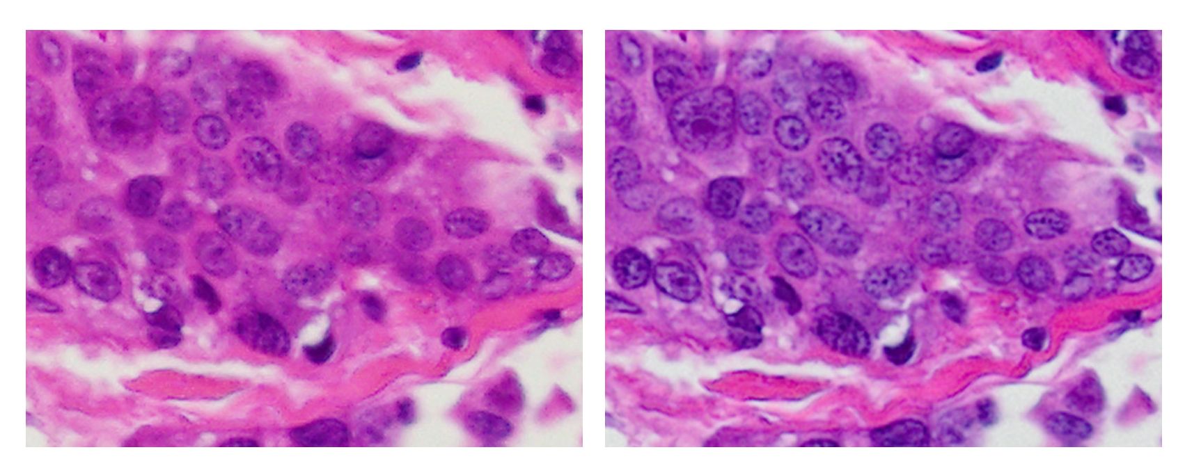
Without OSIA (left) vs. with OSIA (right)
Focus peaking: helps you identify which sample regions are in focus through a visual focusing aid that shows in-focus areas in color and out-of-focus areas in a gray overlay on the live image; this feature is particularly helpful for observing thick specimens
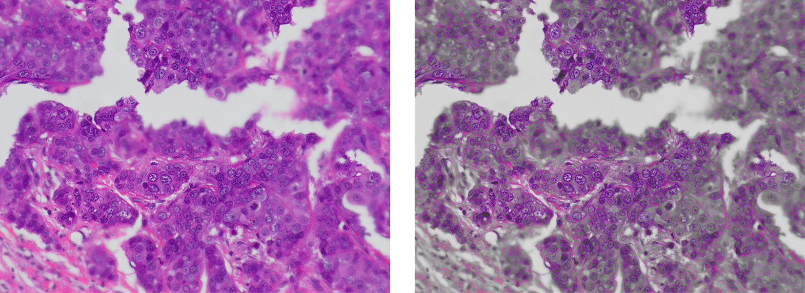
Without focus peaking (left) vs. with focus peaking (right)
High contrast mode: adjusts the exposure time and contrast settings for a higher signal-to-noise ratio—a useful feature for capturing clear images from dim samples in polarization and fluorescence microscopy
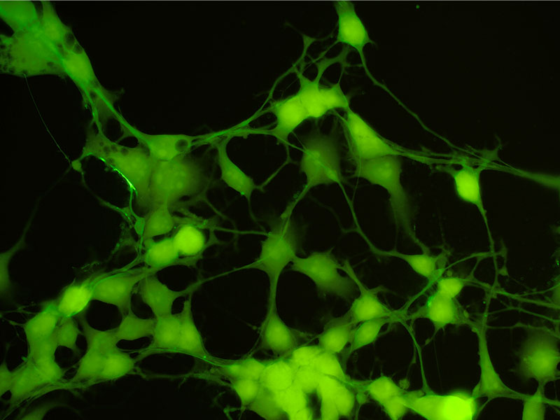
Capture clear images from dim samples with high contrast mode
Fast live: provides a consistently high frame rate even during long-exposure imaging; get smooth images when scanning samples under low light conditions
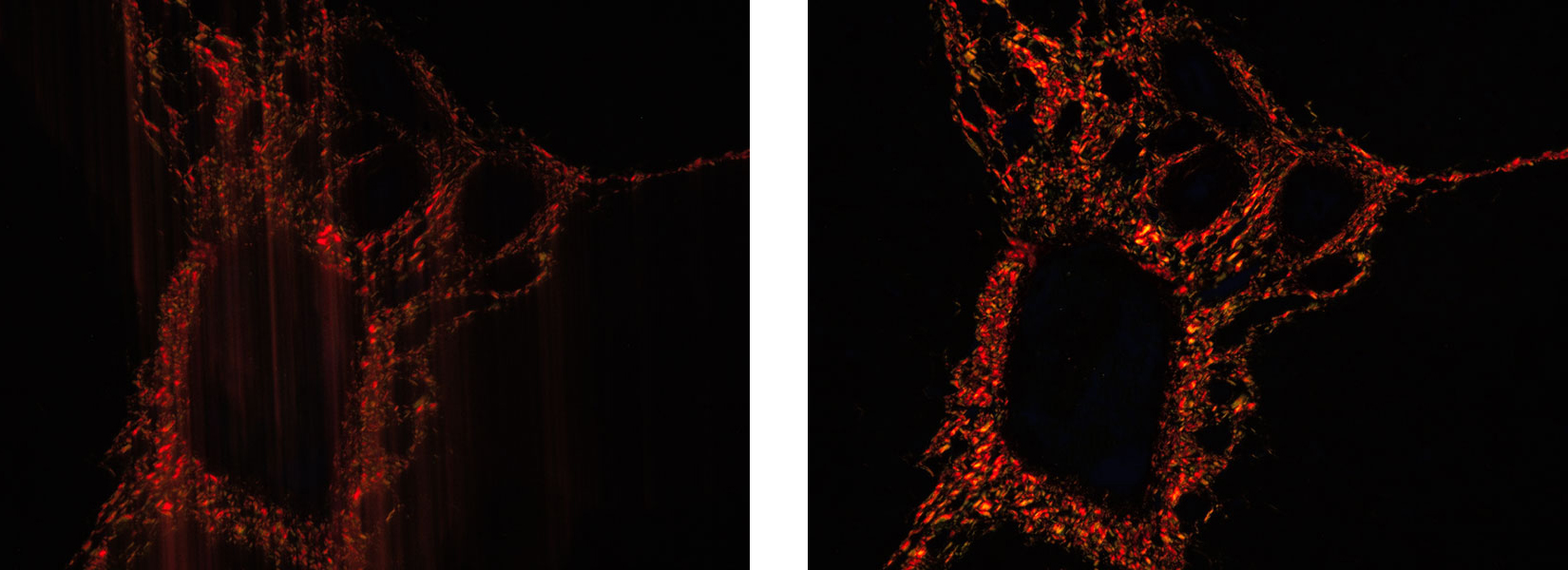
Without fast live (left) vs. with fast live (right)
DP28 or DP23 camera: Which is right for your application?
Now that you know their benefits, you might be wondering which camera is right for your application.
While we recommend comparing camera features and considering how they support your specific application needs, the answer often comes down to a few key features:
- Resolution: The DP28 camera is the right choice if you need a higher resolution. It offers 4K resolution (8.9-megapixels) for efficient collaboration and observation. The DP23, in contrast, provides high resolution (6.4-megapixels) to suit most applications.
- Live imaging: Do you need smooth live images with no distortion or 4K live images for conferences? If so, then our DP28 camera with a global shutter is a good option. Otherwise, the DP23 camera with a rolling shutter provides smooth live images with the detail needed for most applications.
- Sensitivity: The DP28 camera provides more sensitivity for fluorescence applications than the DP23 camera.
If you’re still not sure, use our online camera selector to weigh the options or reach out and chat with an imaging expert. We’re always happy to help!
Related Content
Video: Introduction to the DP23 and DP28 Color Cameras
How to Acquire Microscopy Images that Mimic Human Eye Color Perception