We offer an extensive selection of optical components for equipment integration, delivering solutions to a wide range of fields, including life science research, medical, and industrial. These components include LED light sources, which provide energy efficiency and other notable benefits to microscopy.
In the past, halogen lamps have been used as light sources in microscopes, but now LEDs have become the mainstream. LEDs for microscopes support different lighting methods, such as transmitted illumination and reflected illumination (coaxial illumination).
In this post, learn more about these lighting methods, discover the LEDs that support them, and get tips on how to select a light source for your equipment.
Transmitted vs. Reflected Illumination: What’s the Difference?
If you are designing a microscope device, you may need the instrument to support either transmitted or reflected illumination.
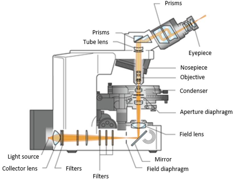
Transmitted illumination (Figure 1) is used with a biological microscope to observe objects that transmit light, such as thin sections of biological tissues, cells, and bacteria. Our BX3M-LEDT is a white LED suited for observation under transmitted illumination.
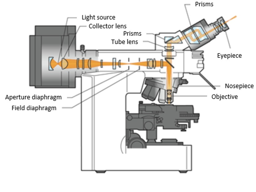
In contrast, reflected illumination (Figure 2) is used for observing objects that reflect light, such as metal surfaces and semiconductors with a metallurgical microscope. Our BX3M-LEDR LED is suited for reflected illumination observation. It is used in many instruments, including Raman spectrophotometers, semiconductor inspection equipment, and 3D measuring machines.
A notable difference between LEDs for transmitted illumination and LEDs for reflected illumination is the size of the diameter of emitted light flux. As the LED for transmitted illumination is used with different types of condensers, it needs a bigger diameter of light flux to illuminate objects evenly. The LED for reflected illumination uses one objective lens to perform the role of both an objective and condenser, so the necessary light flux diameter is smaller. With this design, the numerical aperture (NA) is perfectly matched. This results in the optimal focusing of light and collection of light at the sample plane.
Figure 1. Transmitted illumination |
Figure 2. Reflected illumination |
Comparing Light Sources for Microscopy: LED vs. Halogen
LEDs have many benefits for microscopy. Consider how these light sources compare to halogen lamps. As an example, Table 1 below compares the main features of a 100-watt halogen lamp and white LED for reflected illumination. Even though the LED has the same brightness as the halogen lamp, the LED outperforms halogen in many areas.
100 W Halogen Lamp | Criteria | White LED | ||
|---|---|---|---|---|
| Short | - | Lifespan | ✓ | Long |
| Low | - | Luminous efficiency | ✓ | High |
| Hot | - | Heat | ✓ | Cool |
| Slow | - | Responsiveness | ✓ | Fast |
| Varies depending on the amount of light | - | Color temperature change | ✓ | Almost constant regardless of the amount of light |
| Large | - | Size | ✓ | Small |
| High | - | Color rendering | ✓ | Low |
Lifespan: It is well known that LEDs last a long time. An LED’s lifespan is around 60,000 hours, while a halogen lamp’s lifespan is around 2,000 hours. A longer lifespan means fewer bulb replacements, which saves time, lowers costs, and reduces environmental waste.
Heat: LEDs also have a more stable output than halogen and don’t heat up like halogen. A hot lamp can heat up your workspace and harm some samples. As a cool and stable light source, LEDs enable comfortable, long-term observation.
Responsiveness: The high responsiveness of LEDs lets you immediately begin observation, saving time compared to halogen lamps that take longer to emit bright light.
Color temperature change: Unlike halogen lamps, LEDs maintain a consistent color temperature when you adjust the light intensity. The ability to freely control the light intensity means you can reduce potential damage to the sample from strong light. Consistent color temperature also reduces eye strain, as your eyes don’t have to adapt to the changes in color.
Size: LEDs are generally more compact than halogen lamps, making them easy to integrate even as devices become increasingly miniaturized.
Color Rendering Performance in LEDs vs. Halogen
Note that for general white LEDs, such as BX3M-LEDR or BX3M-LEDT, color rendering is lower than halogen lamps. However, True Color LEDs, such as our U-LHLEDC/U-LHLEDC100 LEDs, have color rendering equivalent to halogen lamps. True Color LEDs are recommended for applications where high color rendering is required, such as observing biological specimens with transmitted illumination.
Spectral Characteristics of a Halogen Lamp and an LED
In transmitted illumination applications, even light intensity across the visible spectrum is also important for accurately reproducing the colors in stained biological samples. Halogens used to outperform LEDs in this area, as the light intensity of general white LEDs can vary at different wavelengths. However, True Color LEDs closely match the spectral characteristics of halogen for accurate color representation.
This performance is illustrated in Figure 3 below, which shows the general spectral characteristics of a halogen lamp with a filter, a True Color LED, and a general white LED.
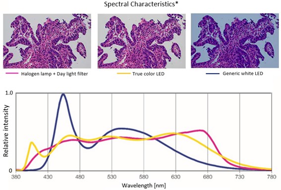
Figure 3. Spectral distributions of a halogen lamp with a filter (left, pink), a True Color LED (center, yellow), and a generic white LED (right, blue).
Learn More about LEDs for Microscope-Based Equipment
When designing new microscope-based equipment, consider integrating an LED for its luminous lighting, energy efficiency, compact size, and other benefits. Our website also provides resources to ease equipment design, such as:
- Datasheets
- 3D CAD data
- RoHS-compatible product lists
To learn more about our high-quality optical components that you can integrate into your microscope designs, check out our OEM resource center.
Related Content
OEM Microscope Components for Integration
How to Minimize an Optical System for a Compact Imaging Device
Direct Access—Download 3D CAD Data of Our Microscope Components