High-resolution whole slide images enable researchers to closely study infection in tissues and cells, making them a valuable tool for researchers looking to better understand how COVID-19 affects the human body.
However, manual slide scanning is a slow process, and researchers need fast results to respond swiftly to the ongoing challenge of COVID-19. As a result, many researchers are turning to automated slide scanners. Read on to learn the key advantages of these systems and how they are used in COVID-19 research.
5 Key Advantages of Automated Slide Scanners for Covid-19 Research
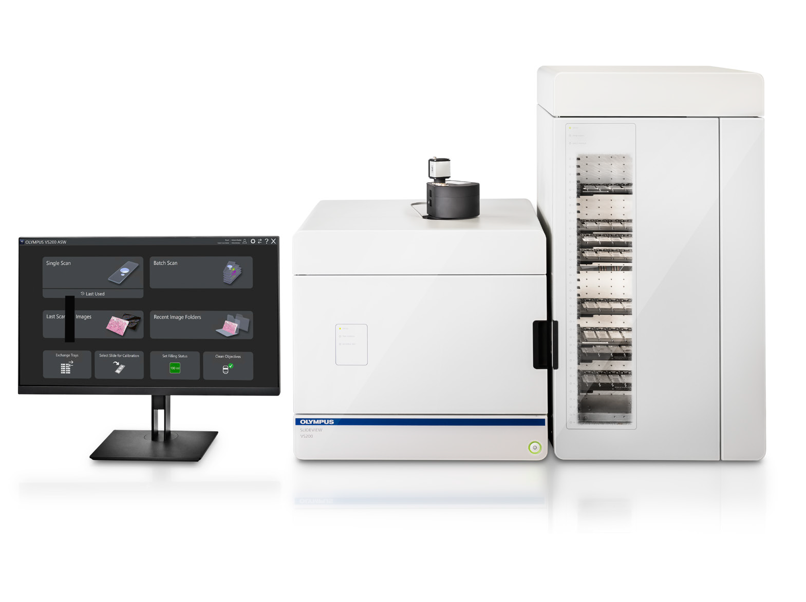
One popular system for COVID-19 research is the SLIDEVIEW™ VS200 research slide scanner. It has a high sample capacity and fast scanning, enabling researchers to capture a large amount of data from a high number of slides in a short amount of time.
Other benefits include:
1. Automated sample detection
The software automatically detects the sample based on contrast, then scans the entire sample area at high magnification with accurate autofocus. This process increases the scanning speed since the system doesn’t waste time scanning the background.
2. High-resolution images for quantification
The system integrates high-performance X Line™ objectives, a light path optimized for the objectives, True Color LED for accurate color reproduction of stained specimens, and uniform illumination. These features enable researchers to acquire large, seamlessly tiled images of whole specimens.
3. Automated immersion oil dispenser
Since the virus is so small, scanning slides using immersion oil can provide high-resolution images (e.g., 60X) to help researchers visualize the infections. The VS200 scanner automatically adds the correct amount of oil to the area you want to scan. To learn how this works, check out our blog post on whole slide imaging with oil immersion media.
4. Flexible imaging modes
The scanner has five different imaging modes—brightfield, fluorescence, darkfield, phase contrast, and polarization. Researchers can choose the mode that works best for their application, and even mix and match methods. For instance, brightfield could be used to visualize tissue, while fluorescence could be used to label COVID RNA.
5. Scan projects
To save time scanning many slides, researchers can save settings in a scan project and then use the same project when running additional samples. This feature is particularly helpful in fluorescence imaging where there are many settings to optimize.
COVID-19 Research Examples Using Automated Slide Scanners
The VS200 slide scanner has proven to be a powerful imaging device to study COVID-19 infections in cells and tissues. The system can be used to visualize and evaluate lung damage from tissue sections obtained from infected subjects (human, mice, etc.) under different conditions. It can also be used to locate and quantify COVID-19 infection in cells or tissue.
Example 1: Imaging lung sections of patients with COVID-19
As an example, Si Wang et al. combined transcriptomics, proteomics, and histopathological technologies to study the lungs of patients with COVID-19. Their findings connect the changes happening in the lung (such as senescence, inflammation, apoptosis, coagulation, and fibrosis) to its underlying cellular and molecular mechanisms. This facilitates the identification of biomarkers and development of treatments for COVID-19.
The researchers used the VS200 slide scanner to capture images of lung sections and performed immunohistology analysis on the images. Figure 6 in the paper shows CD68, a macrophage marker, is labeled for both control and COVID-19 groups. The COVID-19 group shows a larger number of CD68 positive cells, which indicates infiltration of immune cells in the lungs of patients with COVID-19.
Example 2: Imaging lung tissue of mice with COVID-19
In another study, Sarah R. Leist et al. studied multiple aspects of severe COVID-19 disease in standard laboratory mice. The researchers used a VS200 scanner to acquire histological images of mice lung tissue at different days after COVID-19 infection, enabling them to assess the lung damage each day. The researchers also compared the degree of damage between young and old mice, as well as vaccinated and unvaccinated mice. This model facilitates further COVID-19 research on disease severity using mice models.
This study used the RNAscope in situ hybridization assay on tissue sections. In situ hybridization, or ISH, is a type of hybridization that uses a labeled complementary DNA, RNA, or modified nucleic acids strand to localize a specific DNA or RNA sequence in a portion or section of tissue. RNA ISH is used to measure and localize RNAs (mRNAs and miRNAs) within tissue sections, cells, or whole mounts. As a result, the virtual slide images enabled the researchers to measure and map the infection in the tissue sections.
Example 3: Imaging human oral tissue sections
A different study from Ni Huang et al. used the VS200 slide scanner to research COVID-19 infection of the oral cavity and saliva. The researchers scanned RNAscope in situ hybridization samples of human oral tissues using a high-resolution oil immersion objective (60X UPlanXApo, 1.42 NA) combined with the built-in automatic oil dispenser. Thanks to the automated oil dispenser, the researchers could quickly scan the large quantity of high-resolution tissue images needed for the study.
A Powerful Whole Slide Imaging System for COVID-19 Research
As demonstrated in the studies, the VS200 slide scanner enables COVID-19 researchers to quickly acquire high-quality images of tissues and cells for quantification, analysis, and publication. You can learn more about its popular features by reading our post, 5 Ways the VS200 Slide Scanner Can Benefit Your Research.
Related Content
Helping Curb the Spread of COVID-19 with Faster, More Sensitive SARS-CoV-2 Detection
Whole Slide Imaging with Oil Immersion Media: Frequently Asked Questions (FAQs)