On our social media platforms, we love connecting with customers who share an interest in microscopy. One thing we’ve learned over the years is that, for some people, microscopy is one of their life’s passions. And this passion continues no matter your age.
One of our favorite examples is Avalon, an 80-year-old woman from Miramar, Wellington, New Zealand. Her passion is using microscopes to study rotifers.
A Life’s Passion for Microscopy and Studying Rotifers
For those who are unfamiliar, rotifers are microscopic aquatic animals that commonly live in fresh water. These invertebrates are very small—about 0.1 to 0.5 mm long. The term rotifer comes from the Latin word for “wheel-bearer” because they have a circular wheel of cilia that they use to sweep food, such as algae and phytoplankton, into their mouth.
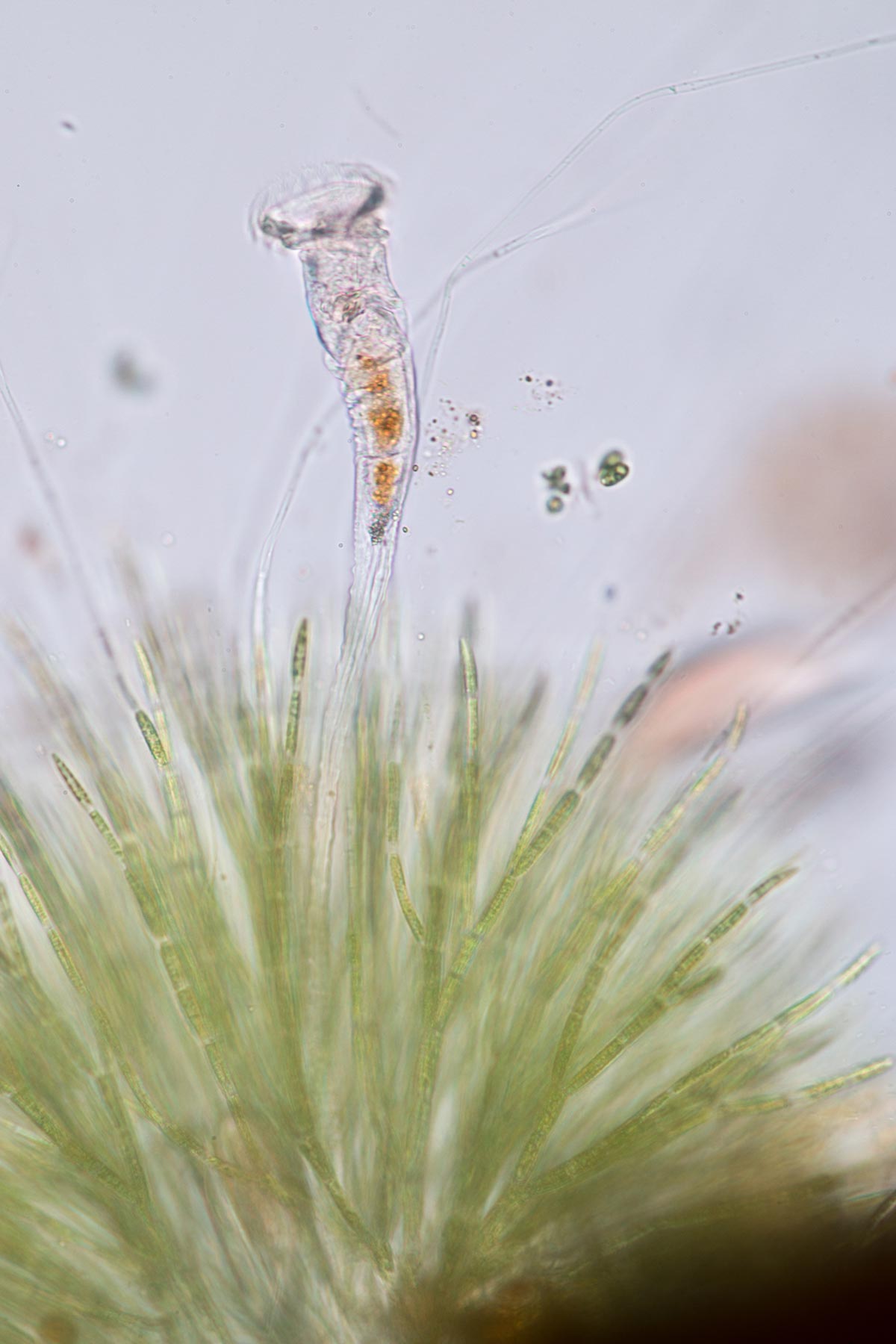
Rotifers are micro-animals that live in fresh water
So, how did Avalon’s passion for studying rotifers begin?
It all started when she was a tech doing water sampling in New Zealand. The health of the rotifers in the water column helps determine the water’s health. If any pollution is in the water, the rotifers will die out. Fascinated by these aquatic animals, Avalon completed a master’s degree specializing in rotifers and continued to study them at a research organization.
Even after she retired, Avalon remained committed to studying rotifers, finding new species, and discovering new information about them. As she began to perform her own informal research, she borrowed an old student microscope from the library and observed rotifers at local botanical gardens.
However, this microscope was too old and simple to give her enough detail to identify the rotifers. To continue her work, she’d need something better.
Finding Her Dream Microscope
Avalon knew she needed a more advanced microscope to clearly see and identify the rotifers. With her goals in mind, she wrote a letter to Olympus to find the ideal microscope.
She wanted to see the rotifer mouth parts at 50X to 100X to identify the rotifer species, so she needed a high-quality research microscope with differential interference contrast (DIC) to get the job done. After meeting with me to discuss her goals and requirements, she discovered her dream microscope—an Olympus BX53 microscope with DIC.
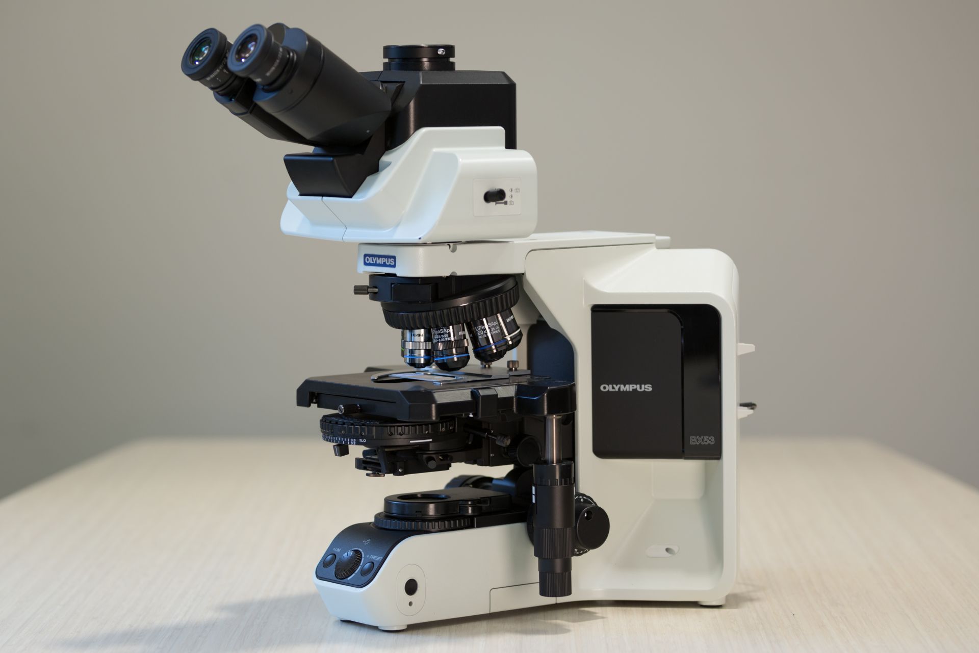
The Olympus BX53 light microscope can be customized for different observation methods
Avalon was determined to get her dream microscope. While it took some time, she was able to achieve her goal and Olympus helped her buy her ideal microscope.
Seeing Rotifers Clearly with High Magnification
Avalon loves using her new BX53 microscope. In particular, she likes the clarity it provides for identifying rotifers and other samples as she hunts for new information.
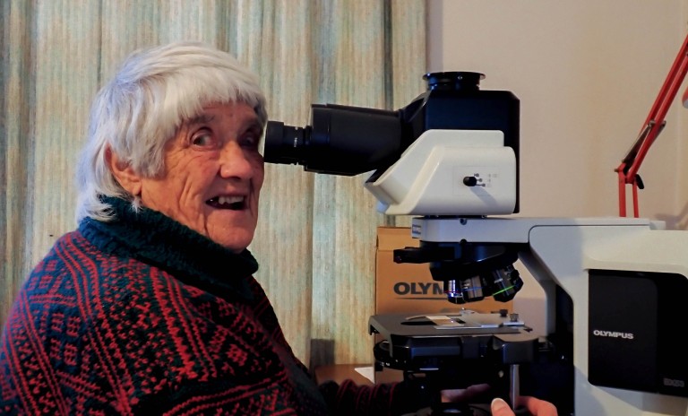
Avalon loves using her new Olympus microscope to identify and study rotifers
Once she collects, counts, and identifies the rotifers of interest, she acid washes them so only the mouth parts remain. Then she looks at the mouth parts at 600X to 1000X magnification to identify the rotifers. While she uses brightfield for the initial scanning, the DIC capability is helpful for identification.
Aside from showing off her Olympus microscope every chance she gets, her next goal is to purchase a microscope camera. While she is new to using cameras and computers, she is determined to learn so that she can share her work.
A Lifelong Connection to the Scientific Community
Avalon stays closely connected to the scientific community. If she discovers a new breakthrough or is unsure about something in her research, she’ll share it with other members of her scientific group. For instance, she once discovered a species of rotifers unknown to be in New Zealand. She will also preserve interesting samples from her nature walks and send them to the community.
To see other interesting samples our customers are researching under the microscope, be sure to follow us on Instagram.
Related Content
Video: BX53 High Luminosity True Color LED
Digitizing Slides Using a Manual Microscope and Digital Camera
A Century of Creation—The Story of Our Life Science Imaging Systems