Need inspiration for our 2022 Global Image of the Year (IOTY) contest? Check out the global winning image from our 2021 competition! This beautiful image of an Arabidopsis thaliana flower, captured by Jan Martinek from the Czech Republic, impressed the jury and took the global prize for masterfully blending artistic creativity with science.
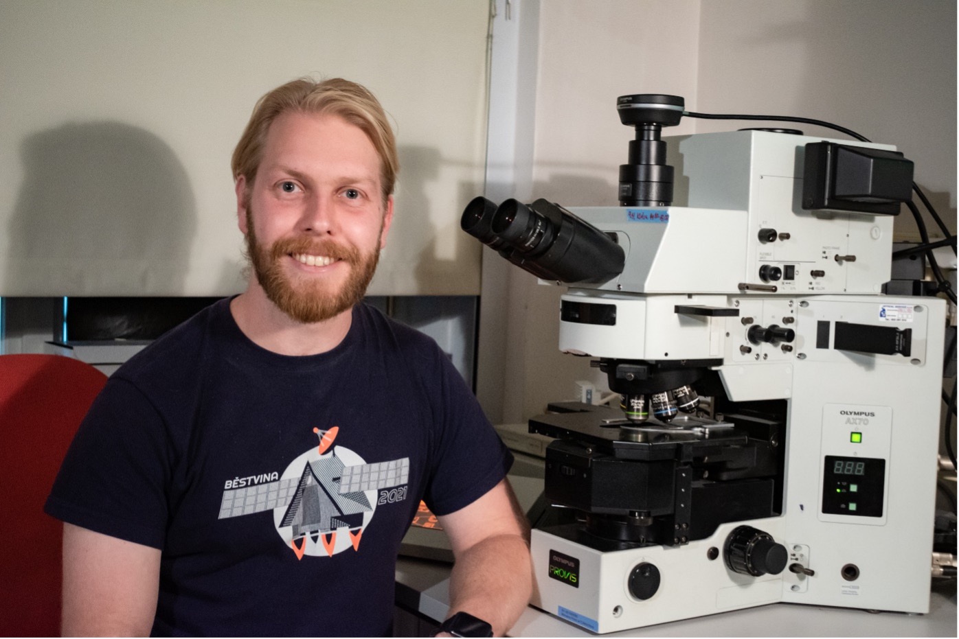
Jan Martinek captured the winning image on an Olympus AX70 system
As part of our annual tradition of sharing the stories behind the winning IOTY images, we chatted with Jan to learn more about the image, sample, and techniques used to capture it. Jan is a PhD student at the Department of Experimental Plant Biology at the Charles University in Prague. He works in the Laboratory of Plant Cell Biology and Biotechnology, where his team focuses on the plant cell cytoskeleton. Microscopy is the main method used in his plant cell research, which led to this stunning capture.
Check out the interview below!
Q: What does your winning image show?
Jan: In this fluorescent microphotography, you can see pollen tubes growing through the pistil of Arabidopsis thaliana. The flower was chemically fixed and cleared to become transparent, then stained with aniline blue. This dye binds to callose, a polysaccharide common in cell walls of pollen tubes, and has green-yellow fluorescence. I did this to measure how efficiently my mutant's pollen fertilizes the ovules.
Q: How did you create this image? Did you face any challenges during the process?
Jan: This picture is captured on the oldest microscope in our lab, an Olympus AX70 Provis microscope. Arabidopsis flowers are quite large, about 2–3 mm, so I used a 4X objective. It is a fluorescence microphotograph in UV excitation light with a wide-pass emission filter.
As for challenges, the samples must be fixed, cleared, softened, and stained to prepare them for microscopy. The protocol is not complicated but rather time consuming.
Q: Why did you choose this image as your entry for the IOTY competition?
Jan: I didn't find the image particularly outstanding myself, but when I uploaded it on my Instagram, it was very successful. So I submitted the image to the competition with other pictures that I found more interesting. My aesthetic taste is often different then the taste of my colleagues and friends, and we often disagree on what is the best picture from the series to upload to my Instagram. I’m glad I listened to the popular vote this time!
Q: What message about the image would you like to share?
Jan: I took this picture together with hundreds of other flowers just to measure the lengths of the pollen tubes. In my research, these measurements will be converted into a graph in Figure 7c, a small piece of data to support my hypothesis. All the microscopic beauty stays hidden behind the results section, so I share these aesthetic by-products of my research on my social media. Nature, and especially plants, are beautiful. And it is great that competitions like yours help to share this message.
Q: Where and when did you first learn to use a microscope?
Jan: I was fascinated by nature since I was a little boy―always catching bugs and butterflies and collecting rocks. My first microscope was basically a toy. There was a science quiz with prizes in a kid’s journal (featuring questions like, how many legs does a spider have?), and the main prize was a toy microscope set. I wanted to win it so bad, and to my surprise, the microscope arrived a week later. Many years later I realized my parents probably bought it for me instead. Later I participated in the Biology Olympiad. Thanks to this, I got into a summer school of biology and chemistry for high school students in Běstvina. Thanks to Professor Jan Černý there, I got my hands on a fluorescence microscope for the first time. It was love at first sight.
Q: When did you become inspired to use microscopes to create art?
Jan: Using my first toy microscope, I tried to capture the images through the eyepiece with my old digital camera (an Olympus C-4000 Zoom). With the vignetting and soft focus of the eyepiece, it usually looked like colorful planets on a black background of space. It was beautiful, and I loved it, but it was more art than a practical scientific illustration.
Today, it's hard not to be inspired when seeing the beauty hidden in the microverse. Plant tissues are especially mesmerizing with their geometric shapes and symmetries. Next to biology, my hobby is landscape photography, so I naturally connected microphotography with fine art photography. I used some tricks about composition or post-processing that I learned in landscape photography in my micro photos.
Q: What do you find most fascinating about microscopy?
Jan: It reveals another level of complexity and beauty even in seemingly dull objects and specimens. I think everybody is fascinated when they discover something previously hidden.
Q: Do you ever perform microscope imaging as a hobby outside of your work?
Jan: My pictures are almost exclusively by-products of my work, and I rarely have a time to do microscopy just for fun. Luckily, my research gives me more than enough opportunities to capture the beauty of the microverse.
Q: What are you currently working on professionally and artistically?
Jan: Currently, I am working on finishing my thesis. It is about the role of the ARP2/3 complex in plant cells.
Q: What is your perception of and experience with Olympus microscopes?
Jan: My first experience with a real fluorescence microscope was on an Olympus microscope, and we still use the old Olympus AX70 system in our lab as the reliable workhorse for general microscopy.
Share Your Best Light Microscopy Images
If you’re inspired by this story, and the stories of our IOTY 2021 regional winners for Asia, the Americas, and EMEA, then consider entering your own light microscopy images (up to 3 images!) into our IOTY 2022 contest. We can’t wait to see what you capture under the microscope.
Related Content
From Tinkerer to Prize Winner: Meet the 2021 Recipient of the IOTY Award for Asia
Celebrating the Global Image of the Year EMEA Winner’s Festive Fungi