Although Olympus has celebrated the beauty and wonder of microscopy imaging with contests in the past, the 2019 Image of the Year (IOTY) competition was the first that went truly global. To ease the difficult job of judging for our expert jurors, the contest is divided into four categories:
- An overall global winner
- And three regional winners:
- Americas,
- Asia-Pacific
- Europe, the Middle East, and Africa (EMEA)
The following is a profile of the 2019 winner for the Americas, Tagide deCarvalho.
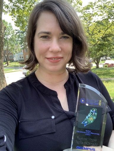
Tagide is the director of the Keith R. Porter Imaging Facility at The University of Maryland, Baltimore County (UMBC) in the United States. There she manages the use of high-end microscopes for research, collaborating with other faculty on a wide range of research topics that span from cell biology to nanoparticle development.
Interview with Tagide deCarvalho
Q: What does your winning image show?
A: This is an image of a live tardigrade, also known as a water bear or moss piglet where you can clearly see the digestive tract from the mouth all the way down to the cloaca. At the front end, you can see that the mouth is a tube armed with stylets used to pierce food such as plant cells or algae and the muscular pharynx is used to suck in the juices.
Q: How did you create the image?
A: The variety of colors in this image stems from a combination of fluorescent dyes and the natural fluorescence of certain tardigrade structures. Live water bears were incubated in calcofluor white and Congo red and imaged on a confocal microscope using a 20X objective lens and three collection channels (blue, green, and red).
Q: What do you find exciting about the image?
A: I like this image because tardigrades look like a “macro” organism, so they have more personality than most microorganisms. And this one looks like it is waving at you!
Q: What do you find most fascinating about microscopy?
A: You never know what you will find.
Q: Where and when did you first learn to use a microscope? Do you have a scientific background?
A: In college, I was originally an art photography major and printed black and white images in my own closet darkroom for many years. I decided to switch to a biology degree but wanted to continue to make art for myself that would incorporate my interest in science. So, I approached a professor in the Department of Anatomy to learn how to do electron microscopy so I could use this technique to produce art. He agreed to hire me as a research assistant because I already knew how to develop and print photographs, which was a significant aspect of microscopy before digital cameras. However, I did not figure out a way to use this form of microscopy to make art but continued to use microscopy during my training as a scientist. And then when I became the KPIF director, I realized that I had the opportunity to finally make this happen.
Q: Where does this fascination stem from?
A: Some of my earliest memories were looking through the microscope at my grandfather’s oncology (cancer) clinic. He published a paper on a microscopy technique called “The Violet Light Microscope: A Method for Visual Estimation of Heme in Living Cells” with Maurice Wilkins, who is a Nobel laureate.
Blurring the Line Between Art and Science
Q: How long have you been creating art with a microscope?
A: I have been creating “sci-art” since I began my current position as the imaging facility director 4 years ago. I consider the non-research micrographs that I take for myself to be “sci-art” for a few reasons. When I am imaging a sample with the intention to make art, I focus on aesthetics (such as composition and color) rather than content. Moreover, I will digitally remove debris from the image during post-processing, which is not acceptable for a research image. However, my images blur the line between art and science because they are informative. For example, I have been approached by several tardigrade experts regarding my technique because they can see structures in the winning image that they have not been able to clearly observe with their current protocols.
Q: Could you tell us about what you are working now?
A: Currently, I am working on my own exciting discovery (using transmission electron microscopy) where it looks like one type of bacteriophage will hitchhike a ride to its bacterial host by attaching to another type of bacteriophage, a phenomenon that has never been observed before.
Olympus Life Science is grateful to all contributors to our global Image of the Year contest, and we can’t wait to see what’s in store for this year’s edition. Stay tuned!
Related Content
Announcing the Winners of Our 2019 Global Image of the Year Award
Olympus Life Science—Instagram
Olympus Digital Image Galleries
Olympus BioScapes – Olympus’ International Digital Imaging Competition from 2004 to 2014