Detection Methods in Confocal Microscopy
Confocal microscopy is a classic technique for examining protein expression and interactions in thick tissue specimens. In contrast to widefield epifluorescence microscopy, which captures emitted photons from the entire field of view (including photons outside the focal plane), confocal microscopy uses a pinhole that is conjugate to the focal plane to reject out-of-focus light and create an optically sectioned image of only the plane of tissue in focus.
The confocal method is effective at obtaining sharp, high-resolution images of thick samples, but the inherent loss of light from the pinhole makes it challenging to capture images with a high signal-to-noise ratio. Because of this, it’s critical to ensure that the rest of the microscope’s optical train is highly efficient to minimize the laser exposure required to generate an image and detect as many of the emitted photons as possible.
Differences Between Filter and Spectral Light Detection Techniques
In filter-based detection, you place a bandpass filter in front of your photomultiplier tube (PMT) to select which wavelengths of light are going to your detector. In spectral-based detection, a grating or a prism is used to create a spectrum of the emitted light, enabling you to finely tune which band or bands of the emission spectrum you wish to detect.
Spectral-based detection also enables you to perform “lambda scans,” in which the entire spectrum of emitted light can be profiled in specific bandwidths and step sizes.
While spectral-based detection methods enable more flexible experiments, there has traditionally been a trade-off in terms of sensitivity. Spectral-based detection provides greater flexibility, but filter-based detection is typically more efficient.
Origins of Spectroscopy in Microscopy
To create a spectrum, spectral detection technology has relied on two methods—reflection-type diffraction gratings and prism-based systems. The reflection technology debuted in the first commercially available confocal microscope in the late 1990s (Figure 1). Shortly thereafter, a prism-based confocal system became available.
As mentioned above, spectral detection methods tend to be less efficient versus filter-based methods. There are three contributing factors to this difference in efficiency:
- Loss of higher order diffractions: When light is reflected off a diffraction grating, multiple orders of diffraction are created. Since only the first order of diffraction progresses through the detection path, the higher orders of diffraction are lost.
- Polarization-dependent loss: The reflectance of light differs depending on the polarization state, with the diffraction efficiency of P-polarized light being less than that of S-polarized light for reflection gratings.
- Wavelength-dependent efficiency of reflection grating: Diffraction efficiency reaches a maximum at a certain wavelength, and the efficiency at higher or lower wavelengths will be worse.
Reflection-type diffraction gratings suffer from all three of these factors and are the least efficient method of spectral detection. Prism-based detection methods overcome the issue of losing higher order diffraction light, but the spectral resolution suffers at longer wavelengths. Because of these drawbacks, neither technology has been able to fully replace the need for filter-based detection systems.
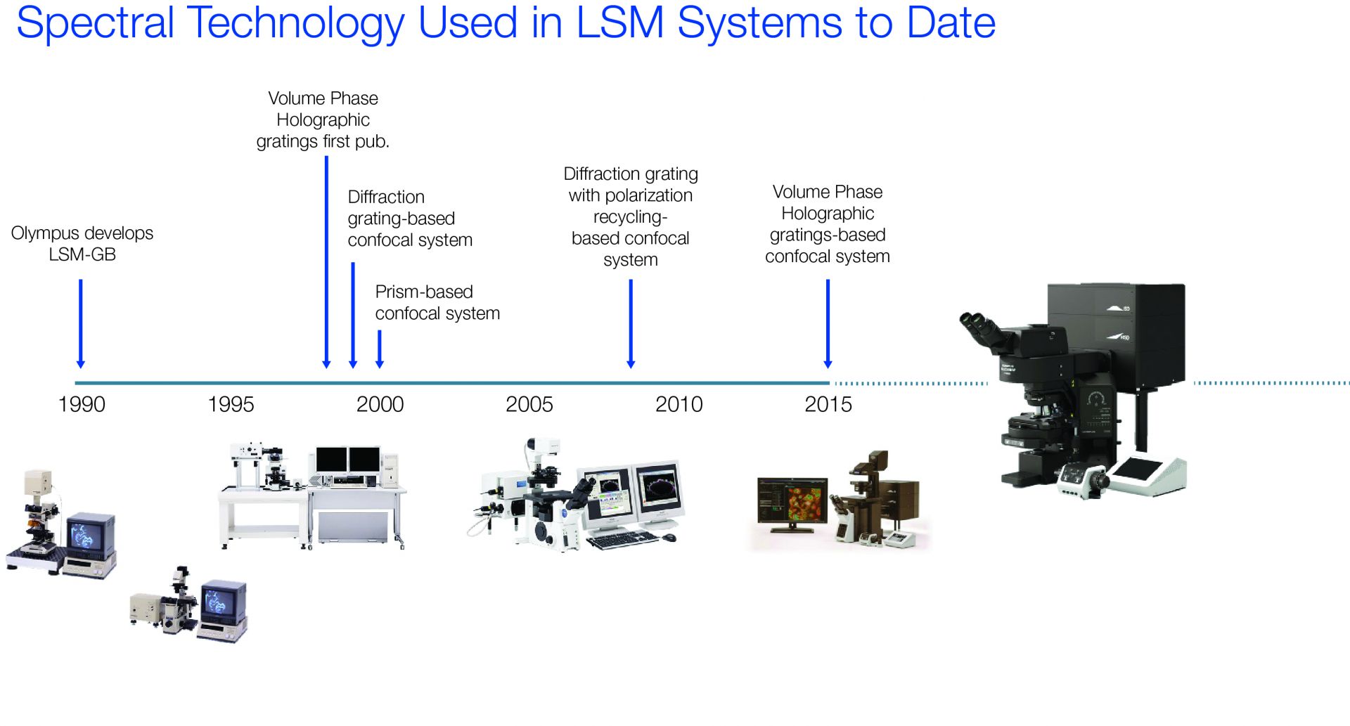
Figure 1: Timeline of confocal microscopy and spectral-based detection technologies
Volume Phase Hologram: A Different Approach to Gratings
For almost two decades, reflection-type diffraction gratings and prism-based technologies powered the majority of spectrology applications in confocal microscopes across all manufacturers. However, microscopy is not the only field to rely on spectroscopy as a means of image formation—astronomy does as well. Spectroscopy in astronomy is similar to that of microscopy in the sense that astronomical objects of interest (such as galaxies and other celestial bodies) also emit light from the ultraviolet to the infrared, and astronomers need to be able to differentiate these components of light to construct accurate images of them (Figure 2).
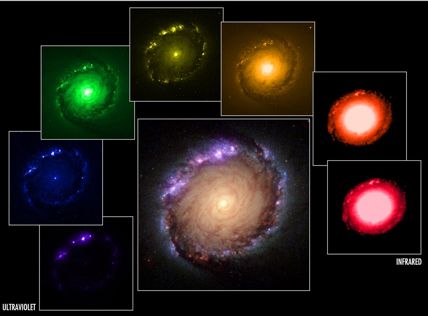
Figure 2: Application of spectroscopy in astronomy to construct an image of a galaxy.
Credit: NASA, ESA, Dan Maoz (Tel Aviv University, Israel, and Columbia University, USA)
One of the key technologies that astronomers use to do this is the volume phase hologram (VPH). Instead of reflecting light off a surface relief grating to produce a spectrum of light, VPH gratings use a transmissive approach, passing light through a grating to diffract the incident light into its spectral components (Figure 3). Astronomy experiments using VPH gratings were first published about 2000, and the technology was rapidly adopted. Very large-scale VPH gratings have since been successfully implemented for spectroscopy in some of the world’s largest laboratories.
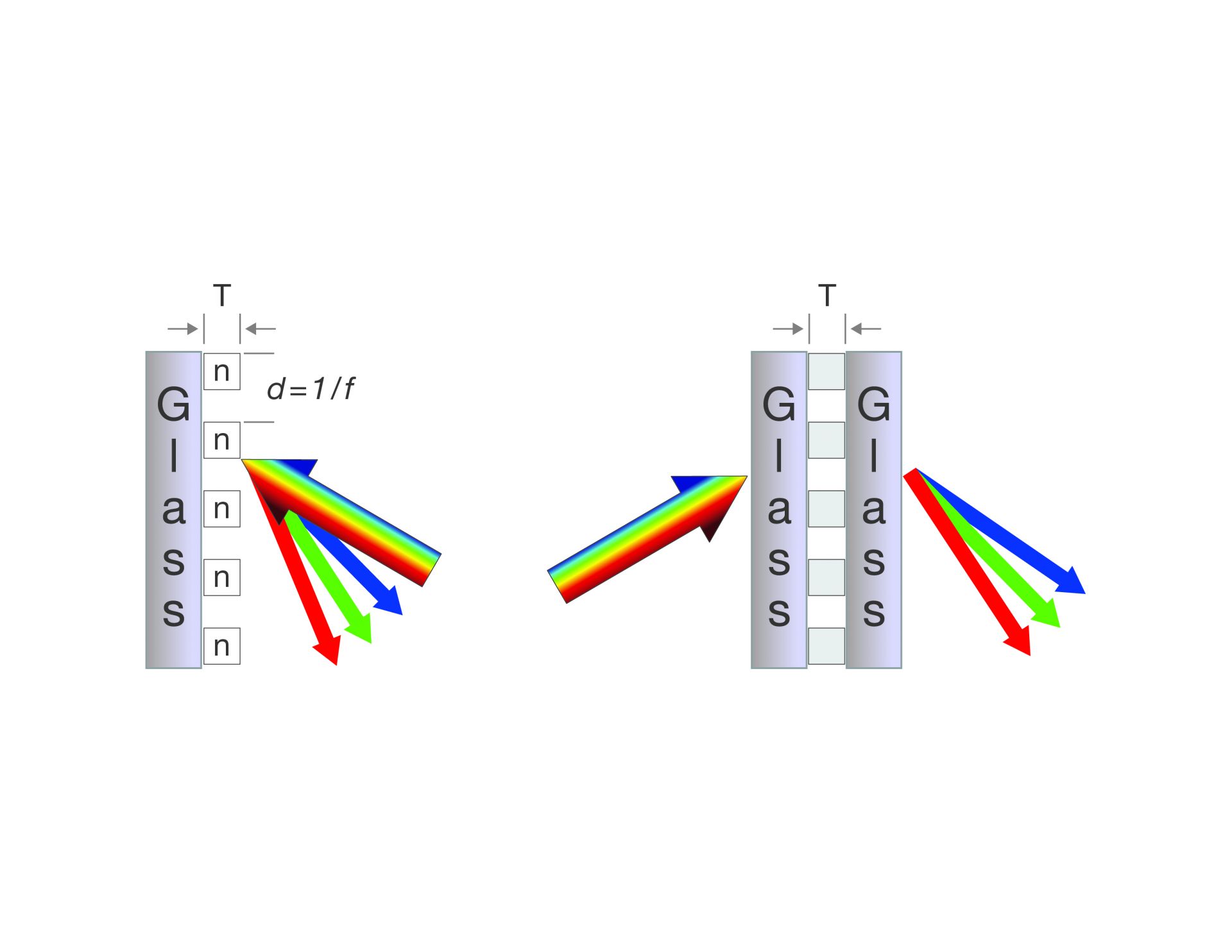
Figure 3: Cross sectional diagrams of the mechanisms of diffraction by surface relief gratings and volume phase holographic gratings. Surface relief gratings (on the left) reflect light off the surface of the grating, whereas VPH gratings (on the right) transmit light through the grating.
In 2016, VPH technology was implemented in a commercial microscope for the first time with the launch of the FLUOVIEW™ FV3000 confocal microscope with TruSpectral™ detection technology, which uses a VPH to enable transmission-type light diffraction. The benefits of using VPH gratings in microscopy include:
- Low polarization sensitivity
- Low scatter (high efficiency)
- High transmission across the spectrum (especially in red compared with reflection-type diffraction gratings)
- More flexible than filters
Because it uses a transmission-type diffraction grating, TruSpectral technology overcomes many of the challenges typically associated with conventional spectral detection methods. For instance, its polarization-dependent loss is minimal compared with the significant loss that typically occurs with reflection-type diffraction gratings. Additionally, the VPH removes the wavelength-dependency of the diffraction efficiency. Reflection-type gratings have a fixed angle so they can only be optimized for one wavelength, whereas the angle of the VPH can be controlled to optimize efficiency at any detection wavelength. This enables higher transmission efficiency across the spectrum, particularly at longer wavelengths.
How VPH Gratings Work in the FV3000 Microscope
Our use of the VPH in our TruSpectral detection technology in the FV3000 confocal microscope relies on three key features (Figure 4):
- A motorized angle adjustment of the VPH to automatically optimize its angle for the wavelengths of light being detected
- An adjustable motorized lambda mirror, which is used to direct specific regions of the spectrum produced by the VPH to the PMT
- A motorized adjustable slit just in front of the PMT, which can be freely adjusted from 1 nm to 100 nm, with 1 nm step sizes in between
Together, these features enable high-resolution linear spectral detection from 400 nm to 800 nm.
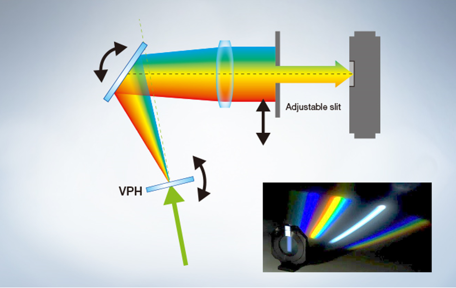
Figure 4: Diagram of VPH-based TruSpectral detection system in the FV3000 confocal microscope
Benefits of TruSpectral Detection for Fluorescence Imaging Using Red Dyes
The VPH provides not only increased flexibility but also significant improvement in the detection system's transmission efficiency, particularly in the desirable red to far-red window (Figure 5).
Red-shifted dyes are becoming more popular for imaging applications since red-shifted light is less phototoxic, enables better depth penetration in tissues, and expands multiplexing capabilities. However, red fluorophores are often difficult to image using traditional spectral-based imaging technologies because of the poor transmission efficiency and spectral resolution of reflection-type diffraction gratings and prisms. VPH-based spectral detection not only enables higher transmission of red light, but it retains the precision and resolution of the resulting spectrum, enabling precise 1 nm spectral resolution all the way out to 800 nm.
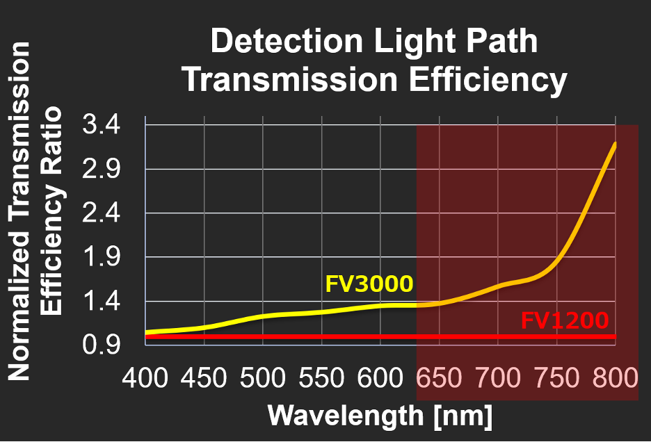
Figure 5: Comparison of transmission efficiency of VPH-based TruSpectral detection technology in the FV3000 microscope versus reflection-based diffraction gratings used in the FV1200 microscope. The use of VPH gratings results in up to 3-fold higher transmission versus traditional spectral detection methods.
The improvements enabled by the VPH grating allowed us to design and build the FV3000 microscope as a fully spectral-based system, meaning that every detector uses the VPH-based TruSpectral detection technology as shown in Figure 4. A fully spectral system with independent channels enables you to unmix multiple signals of varying intensity for simultaneous detection of both bright and dim fluorescence (Figure 6).
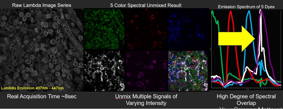
Figure 6: Spectral “lambda scan” of COS-7 cells yields clear separation of highly overlapping signals
From astronomy to microscopy, VPH gratings are proving to be a powerful tool for performing spectroscopy experiments. Stay tuned to see where the voyage takes us next.
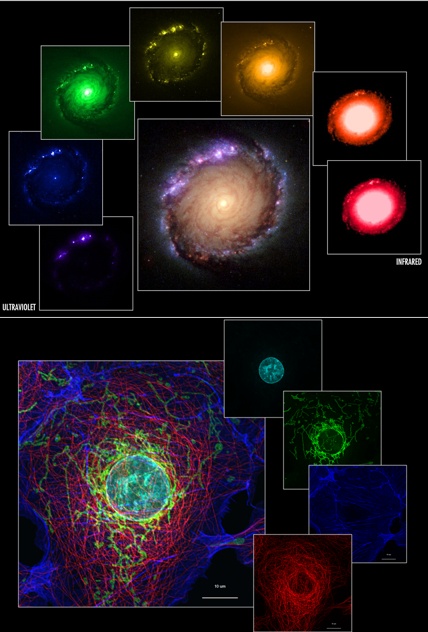
Figure 7: Comparison of spectroscopy applications in astronomy versus microscopy. Top: spectral separation of light emitted from different celestial bodies within a galaxy. Bottom: spectral separation of four different structures within a cell. Top images credit: NASA, ESA, Dan Maoz (Tel Aviv University, Israel, and Columbia University, USA)
Related Content
Multiplexing with the FLUOVIEW FV3000 Confocal Microscope
3D Time-Lapse Imaging of Spheroids with the FLUOVIEW FV3000 Confocal Microscope