Dr. Masayasu Taki is working to elucidate biological phenomena that could not be visualized before through the development of probes based on new fluorescent dyes and near-infrared (NIR) fluorescence. In this interview, we asked Dr. Taki why he focuses on probe development, how the FV3000 Red system helps solve problems encountered in the process of probe development, and what the future holds for NIR imaging.
About Associate Professor Dr. Masayasu Taki
Dr. Taki holds a PhD in Engineering from Osaka University and is currently an associate professor at the Institute of Transformative Bio-Molecules (ITbM) of Nagoya University.
Dr. Taki’s research interests are in the areas of chemical biology, particularly the development of synthetic chemical tools to visualize specific biomolecules as well as biological phenomena using a fluorescence microscope. He has published several papers on long time-lapse imaging through NIR fluorescence and detection of intracellular ions.
Q: What is the current focus of your research?
Dr. Taki: My and my team’s main research is the development of fluorescent dyes for organelle imaging and the development of fluorescent probes from these dyes.
I believe that fluorescent dyes and fluorescent probes have slightly different meanings. Fluorescent dyes are basically things that glow continuously, while fluorescent probes change their fluorescence in response. We have developed fluorescent dyes that have led to the development of probes that can sense events that occur within cells.
| Currently, we are particularly interested in the lipids that make up the organelle. Recently, we reported on the development of a probe that can visualize fatty acid metabolism and its imaging application. Although fluorescent proteins can label membrane proteins, they cannot label the lipid membrane itself. I am researching organic fluorescent dyes because I believe their strengths can be utilized here. And then there is the development of near-infrared fluorescent dyes and probes. NIR imaging is difficult with fluorescent proteins, so fluorescent molecules can help. | 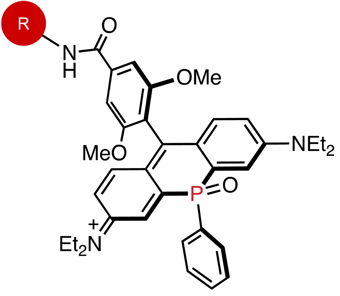 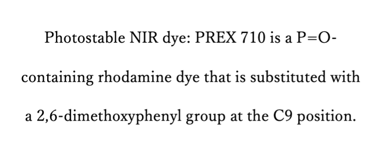 |
Q: Why did you focus on fluorescent dye development and probe development?
Dr. Taki: Fluorescent proteins are powerful tools in fluorescence imaging. However, as I mentioned, the dynamics of lipids and glycans cannot be directly observed with fluorescent proteins. Another problem is that fluorescent proteins tend to fade easily, so it is difficult to continue observation for a long time.
I originally got into imaging while studying at Northwestern University in the US, where lab members were synthesizing zinc fluorescent probes in the laboratory of Professor Thomas V. O'Halloran. It was so beautiful and fascinating to observe the molecules glowing inside the cells that I took the liberty of changing my research theme while the professor was on summer break.
I have been doing fluorescent dye and probe development ever since. For a while, I was doing imaging research on biometals, but when I moved to Nagoya University, I began to wonder “what can only be done with molecules,” and this led me to my current work in lipid dynamics observation and NIR imaging.
Q: How does the FV3000 Red system help you conduct your experiments?
Dr. Taki: One of the NIR fluorochromes we developed is PREX710, which has a fluorescence peak at 740 nm. The sensitivity of the GaAsP PMT detector used in ordinary confocal microscopes drops sharply after 750 nm, so the detector, which is bright enough for ordinary fluorescence observation, is not effective for NIR fluorochromes. However, using the FV3000 Red GaAs PMT detector, NIR fluorescent dyes that were previously difficult to observe can now be observed, which was a nice surprise. I am now reviewing my experiments again because I can observe some things that I had given up on before using the FV3000 Red system.
One of the advantages of using the FV3000 Red NIR solution is the ability to increase the number of observation channels in the near-infrared region. The near-infrared fluorescent dye we are developing can be used with silicon rhodamine (SiR). These are chemical compounds that feature excellent lightfastness and low cell damage due to light irradiation, which makes it possible to observe organelle contacts for a long period of time. The FV3000 Red system’s TruFocus™ Z-drift correction is essential, especially if the observation extends over several hours. The FV3000 Red system’s combination of laser, detection sensitivity, and Z-drift correction is very helpful.
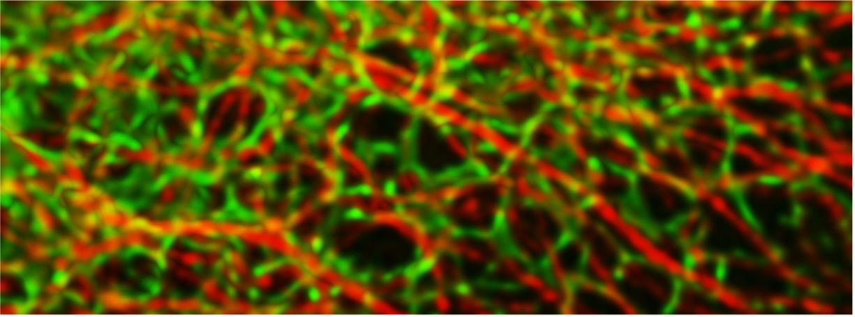
Multicolor imaging of microtubules and endoplasmic reticulum
Q: What is your vision for the future of NIR imaging?
Dr. Taki: I think that most researchers who are currently performing NIR imaging are doing it for deep biological observation. Of course, it is important to demonstrate the usefulness of deep imaging, but, for my part, I would like to develop research that focuses on cellular imaging. Phototoxicity, fluorescence fading, and autofluorescence are some of the challenges faced in cell imaging, and NIR fluorochromes such as PREX710 can overcome these challenges. If we develop a fluorescent dye that is compatible with the 785-nm laser of the FV3000 Red system, near-infrared multicolor observation will become possible.
However, the fluorescence quantum yield of near-infrared fluorescent dyes is not very high, and the main problem is the low luminance of the emission. Low luminance means that the excitation laser power must be stronger, which increases the amount of reactive oxygen species (ROS) and damages cells. This halves the effectiveness of using near-infrared light.
I want to ascertain why they do not glow, and, while overcoming this problem, I would like to develop molecules and create something that researchers around the world would want to use. Also, there is no NIR-compatible lipid dynamics observation probe yet, so I would like to lead the world in developing one. If we develop excellent fluorescent dyes and probes and accumulate examples of NIR imaging at the cellular level, more researchers will use them, and I would like to contribute to the advancement of life science research from the chemical side.
Near-Infrared (NIR) Solutions for Confocal Microscopy
The FV3000 Red system extends the FV3000 microscope’s wavelength detection capabilities to the NIR region. By upgrading each imaging module for NIR detection, including the laser, optics, objective lens, and detector, the FV3000 Red system provides a specialized solution for more sensitive and accurate multicolor NIR imaging.
Learn more about our NIR solutions
References
- A negative-solvatochromic fluorescent probe for visualizing intracellular distributions of fatty acid metabolites
K. Kajiwara, H. Osaki, S. Greßies, K. Kuwata, J-H. Kim, T. Gensch, Y. Sato, F. Glorius, S. Yamaguchi, M. Taki Nat. Commun., 13, 2533 (2022). - Late-stage Functionalisation of Alkyne-modified Phospha-Xanthene Dyes: Lysosomal Imaging Using an OFF-ON-OFF Type of pH Probe
H. Ogasawara, Y. Tanaka, M. Taki, S. Yamaguchi
Chem. Sci., 12, 7902-7907 (2021). - A Highly Photostable Near-Infrared Labeling Agent Based on a Phospha-rhodamine for Long-Term and Deep Imaging
M. Grzybowski, M. Taki, K. Senda, Y. Sato, T. Ariyoshi, Y. Okada, R. Kawakami, T. Imamura, S. Yamaguchi Angew. Chem. Int. Ed., 57, 10137-10141 (2018). - A Highly Photostable Near-Infrared Labeling Agent Based on a Phospha-rhodamine for Long-Term and Deep Imaging
M. Grzybowski, M. Taki, K. Senda, Y. Sato, T. Ariyoshi, Y. Okada, R. Kawakami, T. Imamura, S. Yamaguchi Angew. Chem. Int. Ed., 57, 10137-10141 (2018).
Related Content
Multiplexing and Deep Tissue Imaging with Near-Infrared Confocal Laser Scanning Microscopy (On-Demand Webinar)
Video: Dr. Rebecca Bonfig Introduces the FV3000 Red Near-Infrared (NIR) Solution
Seeing Red: Using Near-Infrared Solutions to Expand the Possibilities of Confocal Microscopy (On-Demand Webinar)