
In many parts of the world, December is a month where folks are focused on the holidays and spending time with friends and family. It’s no surprise that some of the most popular images on the Olympus Life Science Instagram account last month are holiday-related! Check out December’s top 5 below:
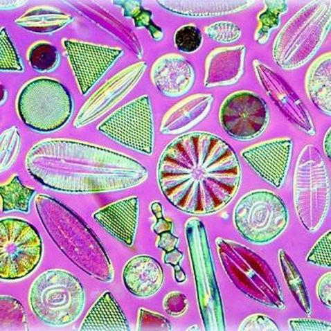
Likely originating during the Jurassic period more than 145 million years ago, diatoms are a major group of algae and one of the most common types of phytoplankton. They're also a popular tool for monitoring environmental conditions (past and present) and are sometimes used to study water quality. As an added benefit, they can create some stunning images!
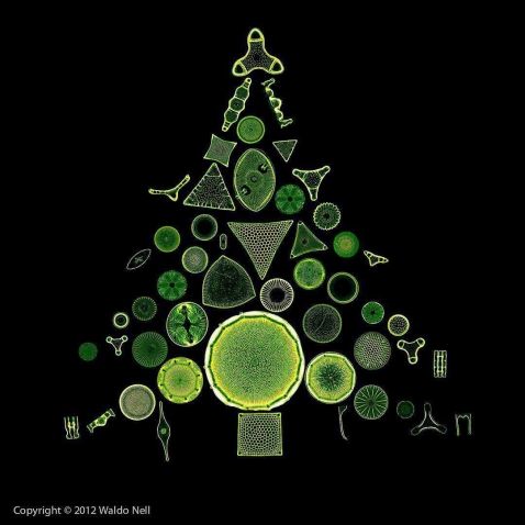
Did you know the average time to grow a 6-foot (1.8 m) evergreen tree is 7 years? The diatoms that make up this ‘tree’ only live for about six days!
Image courtesy of Waldo Nell.
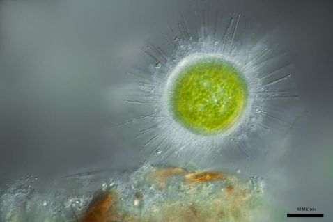
Winter means the cold and flu season, so this brightfield image is appropriately timed. It shows the Acanthocystis turfacea chlorella virus 1, a species of giant double-stranded DNA virus found in the mucous membranes of the human nose.
Image courtesy of Linden Gledhill.
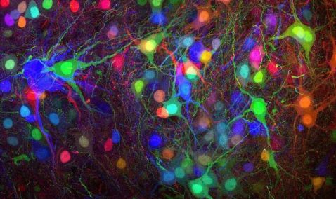
Brainbow is an imaging process that enables individual neurons in the brain to be distinguished from neighboring neurons by using fluorescent proteins. Developed in 2007 by researchers at Harvard University, the technique has since been adapted for use in several organisms, from mice to Drosophila. The neurons and circuits in this stunning Brainbow reminded us of a set of brightly colored holiday lights!
Image courtesy of Dr. Katie Matho.
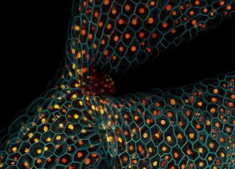
This confocal microscope image shows the expression of different fluorescent proteins in the stem of a thale cress seedling (Arabidopsis thaliana). Also known as mouse-ear cress, it’s a small flowering plant usually considered a weed. A winter annual, this seedling completes its life cycle in just 6 short weeks! Fun fact: Arabidopsis was the first plant to have its genome sequenced.
Image courtesy of Dr. Fernán Federici.
As we enter the new year, we’re thrilled to share the overall top image of 2019:
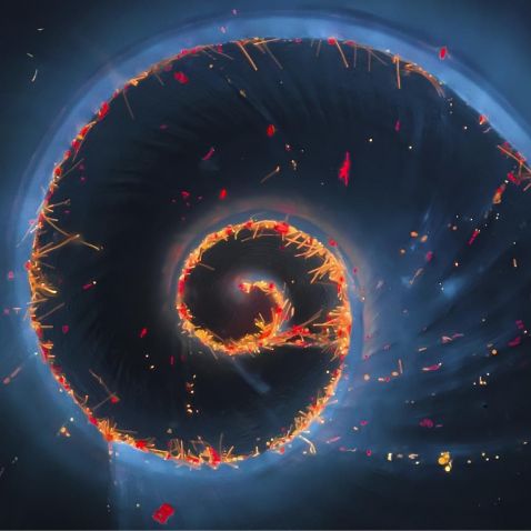
This striking image captured by Håkan Kvarnström is not only a fan favorite but also earned first place in Olympus’ 2018 Image of the Year European Life Science Light Microscopy competition. Captured on an Olympus BX51 fluorescence microscope, this is a small, 2 mm snail found at the bottom of a bottle of water collected from a local fish farm in Stockholm. To learn more about the image, read our recent blog post.
Image courtesy of Håkan Kvarnström.
To see more images like these, be sure to follow us on Instagram at @olympuslifescience!
Interested in sharing your own images?
Visit our image submission site or enter them into our 2019 Global Image of the Year contest.
Related Content
At the Speed of Life—Light Sheet Microscopy Advances for Biological Imaging
