
This month we both dive underwater and travel on land to see the samples captured in our most popular microscope images for December 2021.
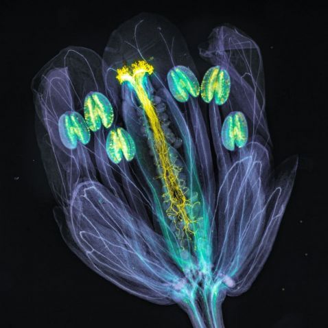
This beautiful image of an Arabidopsis thaliana flower shows the pollen tubes growing through the pistil. The flower tissues were chemically cleared to become transparent, while the pollen tubes were stained with aniline blue (yellow fluorescence) in order to be seen.
Arabidopsis thaliana is useful for genetic mapping and sequencing because of the small size of its genome and because it is a diploid (contains two sets of chromosomes).
Image courtesy of Jan Martinek. Captured with widefield fluorescence microscopy in UV excitation light.
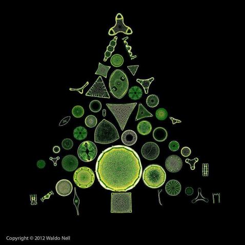
What's the first thing you think of when you hear the word “evergreen”? For many people, it’s a pine tree. Evergreens include many other species, including junipers, hemlocks, redwoods, firs, and even tropical plants, such as palm trees!
Instead of branches and foliage, this “evergreen tree” is actually composed of diatoms that were artfully arranged. For those who are unfamiliar, diatoms are microalgae found around the world in oceans, waterways, and even soil.
Image courtesy of Waldo Nell.
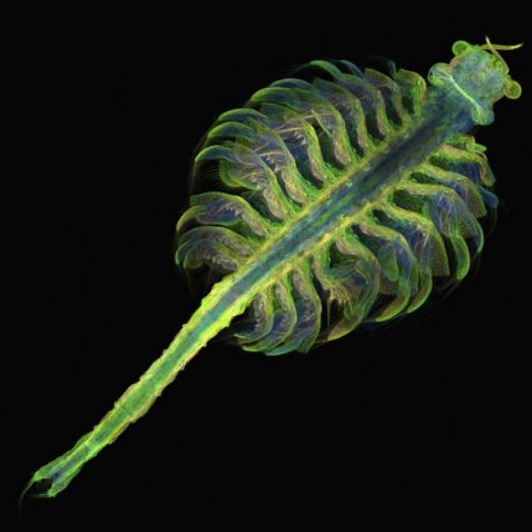
Commonly known as brine shrimp, this is a stunning image of Artemia, a genus of aquatic crustaceans. Able to live in bodies of water with up to 25% salinity, these microorganisms are commonly found in inland saltwater lakes.
Image courtesy of Gopi Shah. Honorable mention in our 2018 Image of the Year competition.
Our 2021 Image of the Year competition is accepting entries through January 31! Learn more at olympus-lifescience.com/ioty.
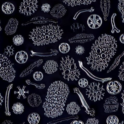
While they may look like edible cucumbers with their long, shapeless body, sea cucumbers are actually echinoderms, which contain an endoskeleton just below the skin. This endoskeleton is formed by calcified structures joined by connective tissue. Here's a close-up look at their various types of bone fragments.
Image courtesy of Yoshihiro Tamaru. 2019 Image of the Year Award submission. Captured using an Olympus BH2 series microscope.
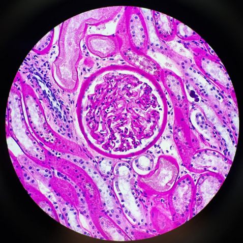
Captured by Kate Murphy, this vibrant image shows a section of a lion’s kidney stained with periodic acid–Schiff (PAS).
Kate explains, “The glomerulus is a loop of capillaries within the kidney, and it’s where the filtration of waste products begins. The small molecules (like water and wastes) will filter out and travel down to the tubules. As it travels down the tubule, important substances are then returned into the bloodstream, leaving the wastes to travel into a collecting duct and eventually be excreted in the urine.”
Image and caption courtesy of Kate Murphy. Captured using an Olympus BX40 microscope.
Bonus video! Because everyone loves tardigrades.
Have you ever taken a close-up look at the claws of a tardigrade? This species, known as the Echiniscus testudo, might not be as resistant as other moss tardigrades but definitely has the coolest claws in the tardigrade universe! Where most tardigrades have hook-like, bent-back claws, Echiniscus brings awesome wolverine claws to the table.
Video courtesy of Benedikt Pleyer. Captured on an Olympus BX53 microscope at 400x magnification with 1000x brightfield.
To see more images like these, be sure to follow us on Instagram at @olympuslifescience.
Interested in sharing your own images? Visit our image submission site or enter our global imaging contest!
Related Content
An Eye for Detail—Our Most Popular Microscope Images for November 2021
Spreading His Wings: IOTY 2020 Asia-Pacific Winner Shares the Beauty of the Microworld
Lifelong Love of the Microscopic—Meet the IOTY 2020 Americas Regional Winner
