
This month we began the new year, but some of our preferred specimens to image under the microscope continue to be old favorites. From Daphnia and tardigrades to plant cross-sections, traditional samples continue to impress with stunning new images.
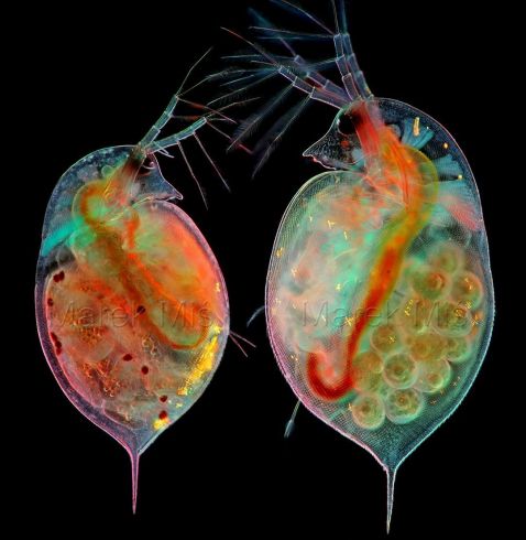
Pictured here are two Daphnia sp. looking deep in conversation. The translucent carapace (upper exoskeleton) lets us view the insides of these microscopic plankton in incredible detail.
Image courtesy of Marek Miś. Captured using an Olympus BH2 microscope in polarized light and darkfield at 100X magnification.
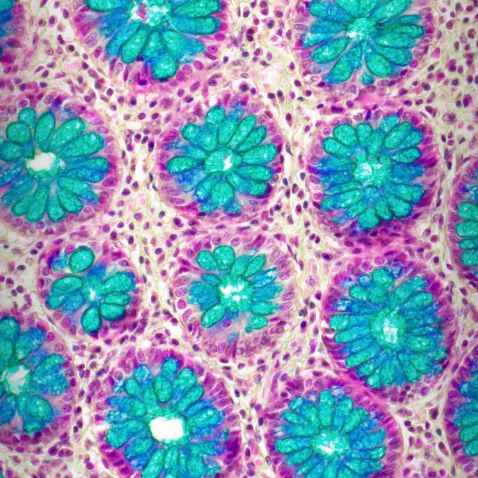
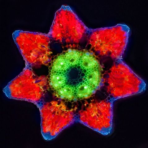
Although this may look like a beautiful flower arrangement, it is an image of cross-sectioned crypts with goblet cells from the colon/large intestine stained with Alcian blue. In contrast to the small intestine, the large intestine has no villi. The surface of the organ is enlarged through crypts. While we know what this specimen is, we loved your comments on what it looks like―wallpaper, a crocheted blanket, or Suzani textiles!
Image captured using a 20X X Line objective.
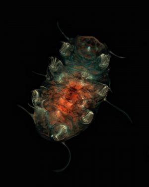
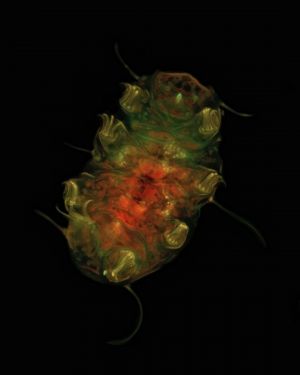
We always love a good tardigrade post! This one looks like it’s saying hello! Luigi Bozzano collected this specimen from moss on an exterior wall of his home in Italy, where he then prepared a slide and viewed it on his microscope.
Image courtesy of Luigi Bozzano. Captured using an Olympus BHB microscope.
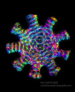
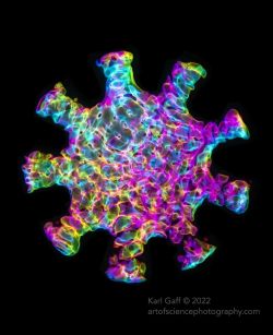
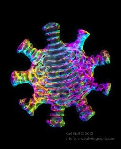
An acoustic mode, known in physics as a phonon, can be thought of as quantized sound waves, much like photons are quantized light waves. Here, Karl Gaff experimented with acoustic modes confined within a virus-shaped cavity to capture the beautiful patterns created by sound.
Image courtesy of Karl Gaff. Captured using an Olympus BX51 microscope.
Captured by Jan Martinek, this cross-section of a horsetail plant shines bright. Jan explains, “This cross-section of horsetail (Equisetum sp.) reminds me of the fake flowers of poinsettia (Euphorbia pulcherrima). I tweaked the white balance and an HSL slider in Lightroom to highlight this similarity, but otherwise, it's just autofluorescence of plant tissues in UV excitation light.”
Image and caption courtesy of Jan Martinek. Captured using an Olympus AX70 microscope.
Fun fact: Jan Martinek was our 2021 Global Image of the Year winner! Check out his winning image and read his interview here. And don’t forget to enter our Global Image of the Year contest for 2022. Visit our IOTY page to submit your entry before the deadline.
Bonus video! When life gives you lemons, throw them under the microscope.
Seen here are “the epidermal cells of the lemon peel (lemon zest). And yes, embedded in the epidermal tissue of the lemon zest you can see stomata. Stomata can be present in other parts of the plant (not just in the leaves!) Stomata are the pores that plants use to take up CO2 and release O2. In other words, stomata are the pores that plants use to 'breathe.'”
Video and caption courtesy of Adolfo Sánchez-Blanco, PhD. Captured using an Olympus CX31 microscope with up to 200X magnification.
To see more images like these, be sure to follow us on Instagram at @olympuslifescience!
Interested in sharing your own images? Enter our Global Image of the Year contest for 2022 before the deadline for the chance to win your own microscope or a set of high-performance objectives. Visit our IOTY page to submit your entry today.
You can also share your favorite microscope images for future posts like this by visiting our image submission site.
Related Content
Diatoms to Brainbows—Our Most Popular Microscope Images for December 2022
Beauty of the Microverse―Meet the IOTY 2021 Global Winner
Moss Piglets to Pine―Our Most Popular Microscope Images for November 2022
