
Spring has sprung, and the world around us is beginning to sprout back to life again. As buds begin to bloom, we love the bold colors that come with the season. Our top images from March boast some spectacular colors as well thanks to staining techniques that highlight sample features.
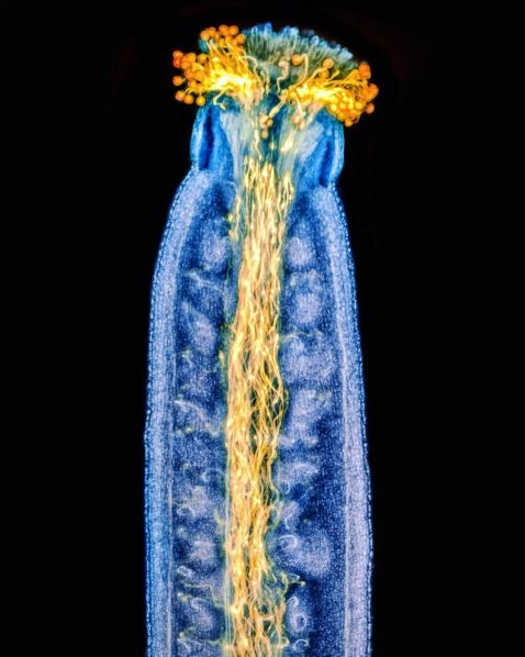
The pollen in the air reminds us spring has arrived. Here you can see pollen tubes growing through the pistil of Arabidopsis thaliana. This spring flower is stained with aniline blue and was imaged using epifluorescence and focus stacking.
Image courtesy of Jan Martinek. Captured using an Olympus AX70 microscope. Fun fact: Jan was our 2021 Image of the Year global winner! Read his IOTY interview here.
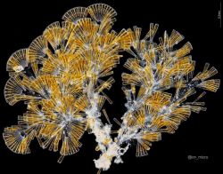
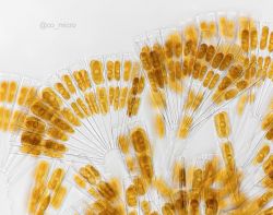
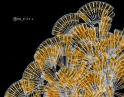
We’re big fans of these stunning diatoms from Sagami Bay, Japan! Diatoms in the genus Licmorphora are found on marine shores and often grow on the sides of other marine or even artificial surfaces. They are considered epiphytes, which are organisms that derive their moisture and nutrients from the air, rain, water, or from debris accumulating around them. This Licmophora diatom arrangement reminds us of beautiful Japanese folding fans!
Image courtesy of @co_micro. Captured using an Olympus BH2 microscope.
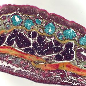
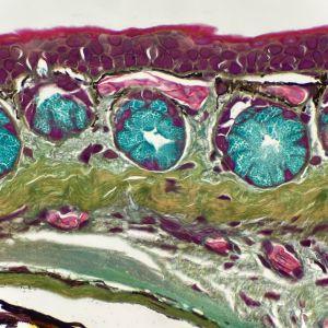
This month we were fortunate to feature another Instagram takeover by one of our favorite histotechs, Kate Murphy. Seen here are colorful images of a salamander’s skin.
“As you can imagine, the skin of amphibians is incredibly unique. The exterior of the skin is a mucosal surface, and it’s always in direct contact with its aquatic environment. This environment is microbially diverse, and so the skin acts as the first line of defense against the pathogens that could be contained in the water. Within this sample you can visualize the presence of multiple mucous glands just under the surface of the skin (bright blue).
The stain we see here is a pentachrome stain. This stain is used to help visualize several different things within the tissue. Elastic fibers are stained black, collagen is yellow, muscle is red, nuclei are dark blue, and mucous is bright blue.”
Image and caption courtesy of Kate Murphy. Captured using an Olympus BX40 microscope.
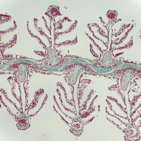
Kate Murphy’s takeover also featured a series of stained organs, including the gills of an eel shown here.
“Gills consist of a single layer of squamous or cuboidal epithelial cells. The primary function of the structures is respiration, and the thin epithelium makes them perfect for gas exchange between the environment and the blood. Along with respiration, the gills are responsible for osmoregulation, pH regulation, and the removal of nitrogenous wastes. Gills are one of my favorite structures to observe under the microscope because they look like little trees!
Gomori’s trichrome, much like the trichrome stain from yesterday, is used to help visualize collagen and muscle within the tissue sample. Except with this stain, the collagen is stained with green rather than blue, and the muscle is still stained with red. Gomori’s trichrome can be applied to all the same tissues and diseases as Masson’s trichrome. It’s a very fun variation of the trichrome stain. I don’t see this method used as often, but I love it!”
Image and caption courtesy of Kate Murphy. Captured using an Olympus BX40 microscope.
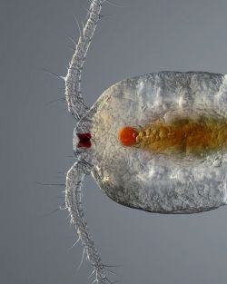
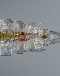

Here’s a tongue twister: this stunning see-through swimmer is a female copepod sampled from a small pond. Meaning "oar feet," copepods are a group of small crustaceans found in nearly every freshwater and saltwater habitat. The one seen here was found in the desert of Al Qudra, Dubai. The sample was imaged at 100X magnification using focus stacking of 76 single shots for the maximum resolution.
Image courtesy of Leonardo Capradossi. Captured using an Olympus UPLFLN 10X objective.
Bonus video! Is it an alien from a distant planet? While it may look otherworldly, this creepy critter is actually an amoeba imaged with phase contrast!
“It has a shell it scoots around in, and on its pseudopodia, you can see the small granules moving in both directions that it uses to help do its amoeba stuff. It’s a stone-cold creep.”
Video and caption courtesy of Katelyn, @dope.microscope. Captured using an Olympus BHS microscope.
To see more images like these, make sure to follow us on Instagram at @olympuslifescience!
Want to share your own images? Visit our image submission site.
Related Content
IOTY Favorites—Our Most Popular Microscope Images for February 2023
All Your Old Favorites―Our Most Popular Microscope Images for January 2023
Diatoms to Brainbows—Our Most Popular Microscope Images for December 2022
