
Our 2021 Image of the Year winners continue to make waves and earn your Instagram likes. From nuclei that look like hearts to hairs splayed out like stars, this month’s top microscope images have us seeing a wide range of shapes!
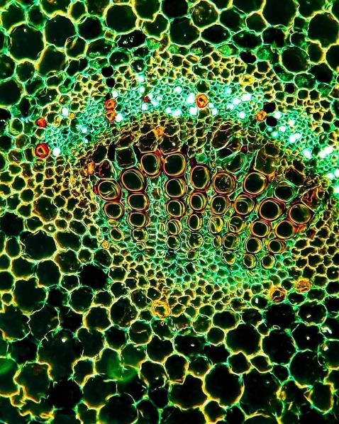
Seen here is the stunning stem of a dicotyledon plant. Dicotyledon plants have distinguishing nodes and internodes, giving rise to leaves, branches, and flowers, contrary to monocotyledon plants. The inner structure of vascular bundles consists of xylem (marked as orange and yellow), phloem (marked as fluorescent green), and multiple fibers.
Image courtesy of Minjun Shin. Captured on an Olympus CX21 microscope at 200X using brightfield microscopy with yellow Rheinberg illumination.
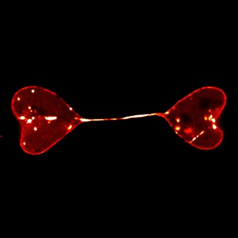
Even cells can have a heart-to-heart moment! Here, the semi-separated nuclei of two cells form a heart-to-heart shape. The nuclei were labeled by lamin, the fibrous protein of the nucleus.
Image courtesy of Di Lu. 2021 Image of the Year (IOTY) honorable mention. Captured on an Olympus FV3000 confocal microscope.
To see the other 2021 IOTY images, visit the Image of the Year page.
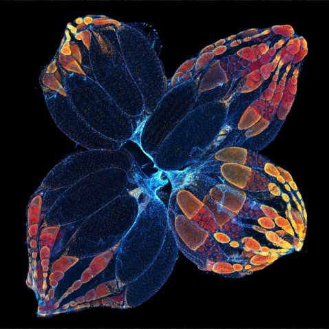
You might not want to find these pesky little critters flying around your kitchen, but they make excellent scientific subjects. Drosophilae, commonly known as fruit flies, are known for their ability to reproduce rapidly. The ovaries, shown here, are not only important but also quite beautiful!
Image courtesy of Yujun Chen. 2021 Image of the Year honorable mention.
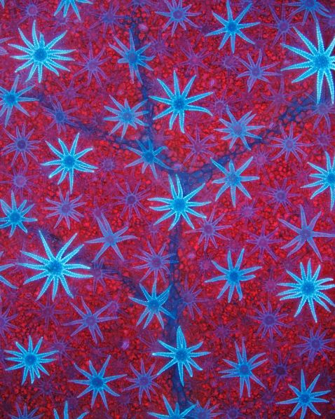
Seeing stars? This image shows the star-shaped defensive hairs covering the surface of a Deutzia leaf, silhouetted against the leaf’s red-fluorescent, chlorophyll-packed cells. A member of the Hydrangea family, the Deutzia plant is a flowering shrub found in Asia, Central America, and Europe.
Image courtesy of David Maitland. 2021 Image of the Year honorable mention. Captured on an Olympus BX51 microscope.
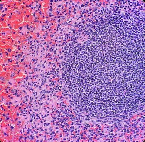
Captured by Kate Murphy, this image is of the spleen of a red-necked wallaby.
Kate explains, “The spleen is the largest organ in our lymphatic system. It primarily stores white blood cells and recycles old red blood cells. It’s essentially the quality control center for our blood. It is possible to survive without a spleen, but it will leave you more prone to infection. Microscopically, we can see the two different 'pulps'. White pulp is the purple, circular clusters of cells. This is the lymphoid tissue. Red pulp (filled with red blood cells) is where blood filtration occurs.”
Image and caption courtesy of Kate Murphy. Captured on an Olympus BX40 microscope.
We all are familiar with the expression that beauty is on the inside. This impressive video shows the epidermal cells of the stem of a flower and the process used to acquire the images, showing the beauty within.
“Embedded in the epidermal tissue, you can also see stomata. Stomata can be present in the stem and in other parts of the plant (not just in the leaves!). Stomata are the pores that plants use to take up CO2 and to release O2. In other words, stomata are the pores that plants use to ‘breathe.’
The liquid I use at the start of the video is distilled water. The distilled water helps maintain the tonicity of the cells in the sample. The water and the cover slip will also enhance the image by avoiding awkward light reflections.”
Video and description courtesy of Adolfo Sánchez-Blanco, PhD. Captured on an Olympus CX31 microscope at up to 400x.
To see more images like these, be sure to follow us on Instagram at @olympuslifescience.
Interested in sharing your own images? Visit our image submission site.
Related Content
Awards Season—Our Most Popular Microscope Images for April 2022
Springtime Green—Our Most Popular Microscope Images for March 2022
Microscopic Valentines—Our Most-Loved Microscope Images for February 2022
