
Spring has turned to summer in our Massachusetts office, and as the warm weather continues, we are seeing more and more microscopic life emerge. Our most popular images from the Olympus Life Science Instagram account again focus on small creatures.
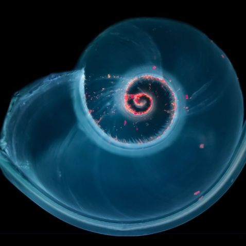
June 20 marked the official first day of summer! Also known as estival solstice, the day is marked by the longest period of daylight and occurs when one of the Earth's poles has its maximum tilt toward the Sun. Summer means beach time for many, and this fluorescence image of a shell has us dreaming of warm days by the water.
Image courtesy of Håkan Kvarnström.
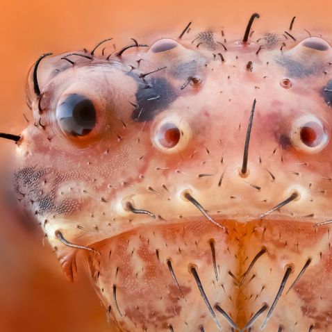
Just a close-up shot of a cute crab spider that is guaranteed to put a smile on your face. Many members of the Thomisidae family are also known as flower spiders, which makes it no surprise that this little fellow was collected from a flower garden in the Netherlands.
Captured on an Olympus BH2 microscope at 10x magnification.
Image courtesy of Marco Jongsma.
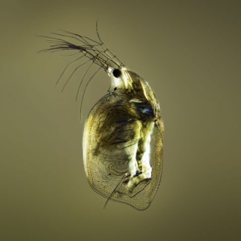
Fun fact: The outer structure of a daphnia, also known as water flea, is so transparent that all internal organs can be seen, including the beating heart!
Image courtesy of Håkan Kvarnström.
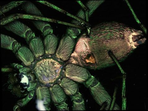
WARNING—This photo is not for arachnophobes or the faint of heart! Get a close-up look under the microscope at the appendages of this spider. Did you know that there are around 40,000 different species of spiders in the world on 6 of the 7 continents?
Captured on an Olympus FLUOVIEW FV3000 confocal microscope with a 10X X Line objective.
Image courtesy of James Lopez, Manager, Olympus Life Science Applications Group.
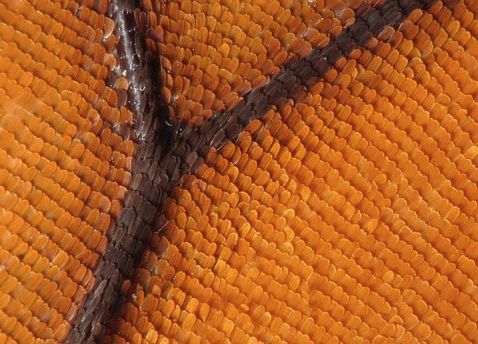
Scientists estimate that there are between 15,000 and 20,000 different species of butterflies in the world. But this one's signature wing pattern always gives it away!
Image courtesy Raul Gonzalez.
Related Videos
Fan favorite Nomadic Nostoc’s “Diatom Trouble, Melosira Edition” is this month’s bonus video.
“All diatoms are faced with the same challenge: their cells get smaller with every generation! Eventually they get so small that it would be impossible to support vital functions and they perish. The reason for this generational size reduction can be found in the structure of their rigid silicon shells (frustules). The name diatom (split in two) is, in fact, a description of their shoe-box-structure in the sense that one half is bigger (epitheca) and can be considered the lid while the other “smaller” half (hypotheca) is the “box”. When diatoms reproduce, they split their two thecae apart and regrow the hypotheca (the old hypotheca becomes an epitheca), which has to be smaller than the old shell halves. As a consequence, diatoms shrink with every division. In order to survive, diatoms have developed a mechanism that kicks in once their size reduction has reached a certain threshold, triggering auxosporolation. The cell bubbles up (auxospore formation) and restores the “original” size of the cell line, which may differ even among the same species of diatom, and regrows a new diatom cell. In the footage, you are witnessing auxospore formation of the filamentous diatom Melosira. Especially beautiful are the star-shaped chloroplasts gathering at the inner surface of the forming sphere, giving these auxospores somewhat the appearance of a Christmas Tree Ball. The time lapse of the spheres took about 3 hours and was very difficult to make, thanks to a swarm of Navicula whizzing around like an angry swarm of bees, constantly displacing the subject.”
Video and caption courtesy of @nomadicnostoc.
To see more images like these, be sure to follow us on Instagram at @olympuslifescience!
Interested in sharing your own images?
Visit our image submission site.
Related Content
Microscope Camera Best Practices for Fluorescence Imaging
