Our SLIDEVIEW VS200 research slide scanner is known for its powerful capabilities that capture high-resolution digital images of whole glass slides.
Beyond its standard features, the system can be enhanced to work with immersion media thanks to an optional liquid dispenser and high-resolution oil or silicone immersion objectives. These features help to maximize image resolution and quality.
Curious to learn how it works? Read on for answers to commonly asked questions about whole slide imaging with immersion media.
How does the VS200 liquid dispenser work?
The slide scanner uses an air pressure pump to dispense a defined volume of droplets onto the slide surface. The number of drops that is dispensed is dynamically dependent on the actual scan area. They are counted via a photo diode droplet counter inside the dispenser head.
This dynamic liquid dispending method is unique to VS200 systems and helps ensure that only the amount of immersion media that is needed is applied to the slide and that no immersion media will drop from the slide into the scanner.
To see the liquid dispenser in action, watch the quick video below.
Related Videos
How does oil immersion media improve slides’ image quality?
Oil immersion microscopy increases the refractive index of a specimen for high optical resolution and image quality. Slides prepared with oil immersion techniques work best under higher magnification where oils increase refraction despite short focal lengths.
What types of oil immersion objectives can be used with the VS200 slide scanner?
For oil immersion in brightfield and fluorescence imaging, three X Line objective options are available: 40X, 60X, and 100X UPLXAPO extended apochromat oil objectives.
How are the different magnifications used in whole slide imaging?
The 40X magnification option offers a large field of view for faster scanning, while the high-magnification objectives (60X and 100X) provide more details at a lower scanning speed.
Here’s a real example of how these magnifications are used. We scanned a human peripheral blood sample (Figure 1 below) of an acute promyelocytic leukemia (APL) using a 40X UPLXAPO oil objective with a 1.4 NA (Figure 2, A and C) and 60X UPLXAPO oil objective with a 1.42 NA (Figure 2, B and D).
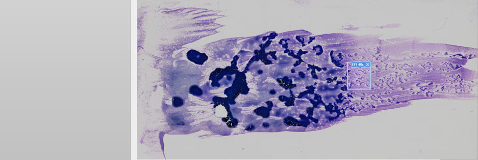
Figure 1. Human peripheral blood sample of an acute promyelocytic leukemia scanned with the VS200 research slide scanner. Images courtesy of Jana Kirsten, Institute for Clinical Chemistry and Laboratory Medicine at the University Medical Center Regensburg.
In this application, the peripheral blood shows the neoplastic promyelocytes with bilobed nuclei or extreme granules (Figure 2, orange arrow). These neoplastic promyelocytes are also found in the bone marrow, as well as myeloid blast cells in which whole bundles of Auer rods (green arrow in A, B, and C) are located. These are important to look for if an APL is suspected.
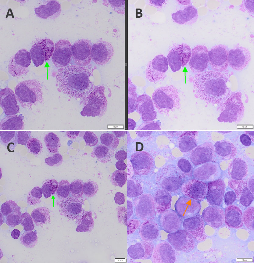
Figure 2: (A) Myeloid blast with Auer rod acquired at 40X magnification but digitally zoomed to 60x. (B) Same blast acquired at 60X magnification and displayed at native resolution. (C) Same image as A but at native 40X resolution. (D) Blast with granules at 60X magnification.
The promyelocytic leukemia (PML) is then confirmed with the evidence of a reciprocal translocation t(t15; 17) via FISH. As a result of this structural chromosomal change, there is a fusion of two genes on chromosome 15 (PML) and chromosome 17 (retinoic acid receptor, alpha [RARα]). Patients suffering from PML can be treated with all-trans retinoic acid (ATRA).
Comparing image A (40X) with B (60X) in Figure 2 shows the benefit of the 40X UPLXAPO oil objective with a 1.4 NA, as the Auer rods and granules are still clearly visible at this lower magnification. Also, the scanning time with the 40X objective was 50% faster compared to the 60X objective (scan time for a 1 × 1 cm area: 40X = 4:40 minutes, 60X = 10:30 minutes).
How do X Line objectives improve whole slide imaging?
All X Line objectives provide:
- Expanded flatness for uniform quality
- Enhanced chromatic correction for exceptional color reproducibility
- Improved numerical aperture for excellent image quality
What types of silicone immersion objectives can be used with the VS200 slide scanner?
For silicone immersion media, three options are available: 40X, 60X, and 100X UPLSAPO super apochromat silicone oil objectives.
How do silicone immersion objectives benefit whole slide imaging?
Silicone immersion objectives offer both a high numerical aperture and long working distance, enabling stable, long-term, high-resolution imaging that is unaffected by changes in temperature.
Related Content
Brochure: VS200 Research Slide Scanner
From Multimode Imaging to Multiplexing: Your Essential Guide to Extracting Rich Data