Life Science Solutions
Optical Sectioning Solutions for 3D Cancer Cell Analysis
Sorry, this page is not
available in your country.
| Select Language for Download |
|---|
With anti-cancer drug approval rates now falling below 5%, the field has begun to adopt new methods for cultivation of cell lines
in vitro to improve the predictive value of cytotoxic agents in vivo.
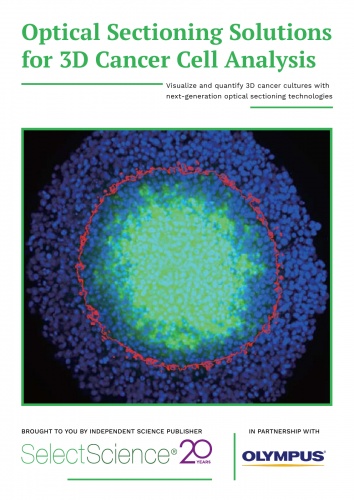
For example, spheroids are an extremely useful tool for modeling the complexity and heterogeneity of tumors in their native environment. However, significant challenges still exist in their analyses, owing largely to the requirement for highly powerful optical lasers for deeper tissue resolution, a type of imaging termed ‘optical sectioning’. If cells are exposed to these powerful light sources for an extended period, they will suffer phototoxic damage, thus limiting one’s ability to conduct continuous analysis for real-time, kinetic data.
In this eBook, we present several cancer-based examples of non-damaging, high-resolution, live-cell and fixed-cell assays, using some of the most innovative optical sectioning technologies. These include the analysis of antibody-dependent cell-mediated toxicity (ADCC), 3D in-gel invasion, DNA damage signaling, mitochondrial uncoupling, and drug efficacy in multicellular models.
Not Available in Your Country
Sorry, this page is not
available in your country.
You are being redirected to our local site.