Automated Organoid Imaging and
High-Throughput 3D Analysis
Discover a smart imaging system that simplifies your workflow,
enabling rapid detection and automated sequential low-to-high magnification
imaging of 3D objects.
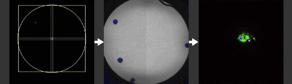
Automated Macro-to-Micro Imaging
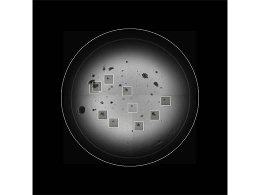
When imaging 3D specimens such as organoids or thick tissue samples, locating target objects can be a challenging and slow process. This task is especially time consuming in a multiple-chambered vessel or multiwell plates. Using this automated acquisition process, you can dramatically reduce the time you spend operating the microscope:
- The FV4000 confocal laser scanning microscope automatically captures images at low magnification.
- Then the FV software’s Macro-to-Micro module detects your objects of interest in the vessel or well and captures them at high magnification.
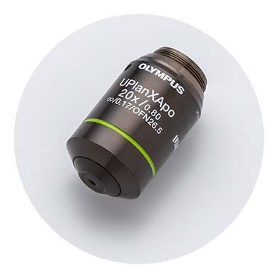
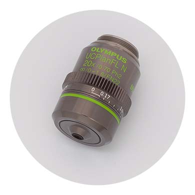
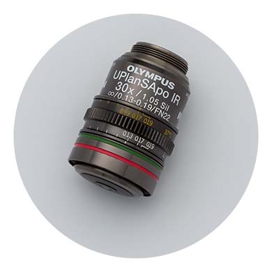
| High-Quality Images with X Line and A Line ObjectivesOlympus offers several objectives with features that help solve the challenges of organoid imaging conditions and observation requirements. For organoids on a glass- or film-bottomed microplate, we recommend the UPLXAPO20X objective from the award-winning X Line™ series. This 20X objective provides an excellent balance of resolution, field of view, and depth of focus. It features a high NA of 0.8, a 600 µm working distance, and air immersion, which makes it easy to image across a microplate. For deep-tissue imaging applications, we recommend the UPLSAPO30XS 30X silicone oil immersion objective, which can reach up to 800 µm deep into your sample. Offering an impressive working distance range of 800 µm to 1800 µm, our UCPLFLN20XPH objective also enables imaging through plastic-bottomed vessels. |
|---|
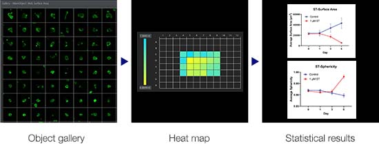
High-Throughput 3D Image AnalysisNoviSight™ 3D cell analysis software is optimized to analyze 3D models on microplates. Once target objects are detected in 3D, you can examine the data using different dimensional views, including:
This flexibility enables you to evaluate the experiment results with high throughput and in a quantitative manner. |
|
| “Automatic organoid imaging enables researchers to perform targeted imaging, which allows for fast detection and sequential high-magnification imaging of 3D objects. This ultimately contributes to the efficiency of our imaging workflow to examine phenotypic changes of patient-derived organoids in response to drug perturbations. Seungil Kim, PhD, Microscopy Team Manager |

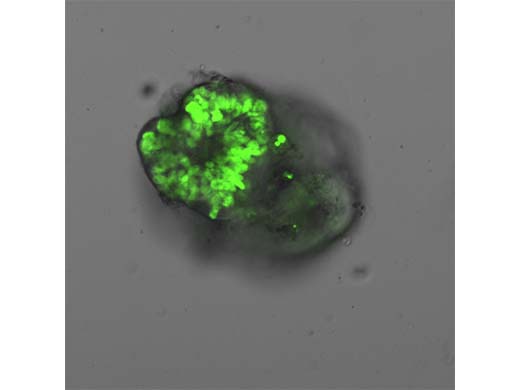
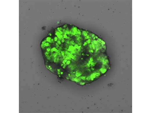
Map at 4X |
Single slice at 20X |
Maximum intensity projection at 20X |
Related Products
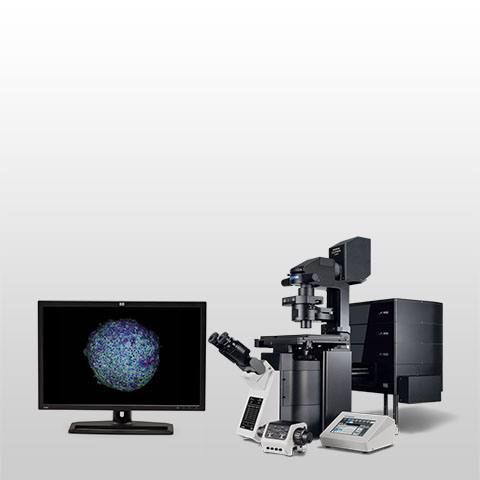
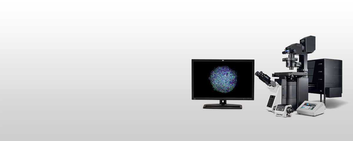
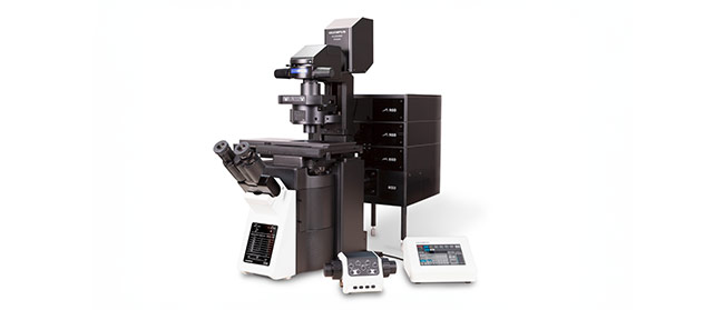
Confocal Laser Scanning Microscope FV4000
*As of October 2023. |
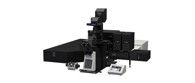
Multiphoton Laser Scanning Microscope FV4000MPE
|
3D Cell Analysis Software NoviSightNoviSight 3D cell analysis software provides statistical data for spheroids and 3D objects in microplate-based experiments. Use it to quantify cell activity in 3D, easily capture rare cell events, obtain accurate cell counts, and improve detection sensitivity. NoviSight software works with a range of imaging techniques, including point-scan confocal imaging, two-photon imaging, spinning disk confocal imaging, and super resolution live cell imaging.
|
Sorry, this page is not
available in your country.
Sorry, this page is not
available in your country.