Multiphoton Excitation ObjectivesEngineered to achieve optimum performance with multiphoton excitation imaging (MPE), A Line MPE objectives enable high-precision imaging of biological specimens to a depth of up to 8 mm for in vivo and transparent samples. | 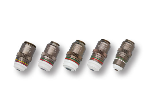 |
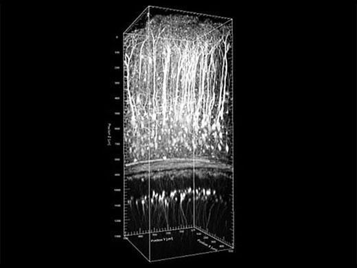 This example shows a Z–stack image of an in vivo mouse under anesthesia from the brain surface to the radiate layer of the hippocampus (CA1) Sample: Thy1–YFP H line 8 week old male Excitation wavelength: 960 nm | Deep Mouse Brain ImagingDeep in vivo, multiphoton brain imaging and optogenetics at high resolution require objectives that have high transmission of infrared (IR) light, a high numerical aperture (NA), and the ability to correct for the depth and scattering of tissue. This objective delivers ultra-broad IR transmission, enabling optogenetic stimulation with visible light down to 400 nm and IR imaging or stimulation beyond 1600 nm. The correction collar reduces the excitation volume, enabling stimulation of single cells or dendritic spines. Combined with the powerful and precise scanning capabilities of the FVMPE–RS microscope, the XLPLN25XWMP2 objective is the right tool for high-precision multiphoton imaging. Image data courtesy of Katsuya Ozawa and Hajime Hirase, Neuron–Glia Circuitry, RIKEN Brain Science Institute, Japan |
|---|
Deep Observation of Fixed Transparent Specimens with Multiphoton Objectives to a Depth of 8 mmOur multiphoton objectives help facilitate breakthrough research on brain function and other vital organs by enabling researchers to see as deep as 8 mm without slicing. The XLPLN25XSVMP2 and XLSLPLN25XSVMP2 objectives support many clearing reagents. Whole Mouse Brain Imaging (XLPLN10XSVMP)The objectives offer a wide field of view with 10X magnification, single-cell resolution with an NA of 1.0, and observations down to 8 mm. The objectives match a wide range of clearing reagent refractive indices (ne: 1.33 to 1.52). Image data courtesy of Hiroshi Hama, Atsushi Miyawaki, RIKEN Brain Science Institute Laboratory for Cell Function Dynamics | 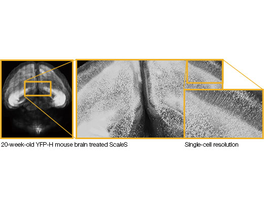 |
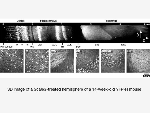 | High-Resolution Deep Brain Imaging of a ScaleS-Treated Mouse Brain (XLSLPLN25XGMP)With a numerical aperture of 1.0 and an 8 mm working distance, the objectives enable deep, high-resolution imaging with a refractive index that matches that of many clearing reagents (ne: 1.41 to 1.52). A maximum intensity projection image (top). Six XY images at different Z positions (bottom). WM: white matter; GCL: granule cell layer, Hil: hilus, LHb: lateral habenular nucleus, MDC: mediodorsal thalamic nucleusImage data courtesy of Hiroshi Hama, Atsushi Miyawaki, RIKEN Brain Science Institute Laboratory for Cell Function DynamicsReference: Nat Neurosci. 2015 Oct; 18 (10): 1518–29. doi: 10.1038/nn.4107. Epub 2015 Sep 14. |
|---|
A Line MPE Objectives Selection Guide
|
Working Distance
(mm) | Magnification | Objective Field Number* | Numerical Aperture | Immersion | Sample | Purpose | |
|---|---|---|---|---|---|---|---|
| XLPLN10XSVMP | 8 | 10X | 18 | 0.60 | Water to oil (ne: 1.33 to 1.52) | In vivo and cleared samples | Wide field of view observation |
| XLSLPLN25XGMP | 8 | 25X | 18 | 1.00 | Silicone oil to oil (ne: 1.41 to 1.52) | Cleared samples | High–resolution observation |
| XLSLPLN25XSVMP2 | 8 | 25X | 18 | 0.95 | Water to silicone oil (ne: 1.33 to 1.41) | In vivo and cleared samples | |
| XLPLN25XSVMP2 | 4 | 25X | 18 | 1.00 | Water to silicone oil (ne: 1.33 to 1.41) | In vivo and cleared samples | |
| XLPLN25XWMP2 | 2 | 25X | 18 | 1.05 | Water | In vivo |
*Maximum field number observable through eyepiece.
MPE Objectives for Inverted Microscopes
|
Working Distance
(mm) | Magnification | Objective Field Number* | Numerical Aperture | Immersion | Applications | |
|---|---|---|---|---|---|---|
| UPLSAPO30XSIR | 0.8 | 30X | 22 | 1.05 | Silicone oil | MPE imaging in deep tissue with a wider field of view |
*Maximum field number observable through eyepiece.
Maximize Resolution in Deep ImagingOur TruResolution objectives maximize resolution and contrast for 3D imaging deep within thick specimens. The objectives are equipped with a motorized correction collar that can automatically and dynamically compensate for spherical aberration while maintaining the focus position. | Related Videos |
TruResolution Objectives
MPE Dedicated Objectives Equipped with an Automated Spherical Aberration Compensation Function
|
Working Distance
(mm) | Magnification | Objective Field Number* | Numerical Aperture | Immersion | Sample | Purpose | |
|---|---|---|---|---|---|---|---|
| FV30AC10SV | 8 | 10X | 18 | 0.60 | Water to oil (ne:1.33 to 1.52) | In vivo and cleared samples | Wide field of view observation |
| FV30AC25W | 2 | 25X | 18 | 1.05 | Water | In vivo | High-resolution observation |
*Maximum field number observable through eyepiece.
Related Products
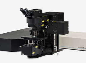
FVMPE-RS
|
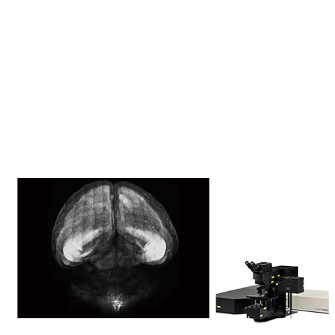
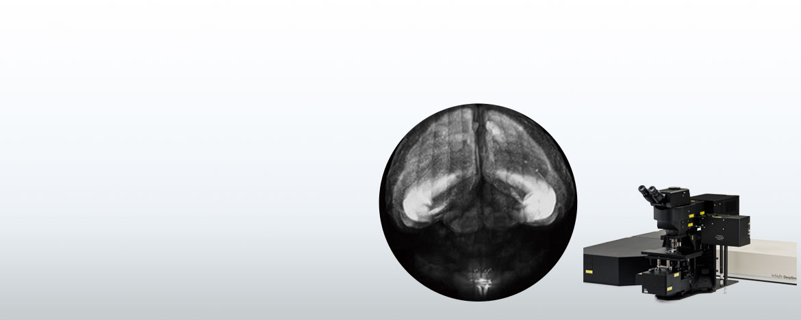
*Banner Image: 20 week old YFP–H mouse brain treated with ScaleS
By courtesy of Hiroshi Hama, Atsushi Miyawaki, RIKEN Brain Science Institute Laboratory for Cell Function Dynamics
Sorry, this page is not
available in your country.
Sorry, this page is not
available in your country.