The amino acid sequence that comprises actin (a highly conserved protein) in humans is very similar to that found in yeasts and plants. Actin monomers bond readily to each other forming two parallel polar filaments arranged into a helical superstructure. The consequence of this structural polarity is that the two ends polymerize and depolymerize at different rates depending on which portion of the actin monomer’s molecular surface is exposed. Assembling in the middle section first, the actin filament grows out linearly from both ends from a central nucleating oligomer. The two polar ends of an actin filament are referred to as the “barbed” and “pointed” ends, with the barbed end polymerizing faster than the pointed end. In the digital video presented above, African water mongoose fibroblast cells (APM line) are expressing a fusion of enhanced yellow fluorescent protein (EYFP) to human beta-actin.
Video 1 - Run Time: 16 Seconds
Video 2- Run Time: 61 Seconds
In order for cell motility to occur, a number of mechanisms must become involved, including actin filaments which play a key role in cell movement during the extrusion of pseudopodia. A pseudopodium, or “false foot,” is a protrusion of continually assembling and disassembling actin filaments that extends and contracts to propel the cell along. Myosin is involved with motility in the trailing end of the cell, causing it to contract by shuffling the cellular fluid to the front and into the pseudopodium, thus breaking the actin filament network. The pseudopodium then extrudes out from the cell body while the filament network rebuilds itself only to be contracted and broken again by the myosin. Many cells, including white blood cells, travel by extending pseudopodia, a phenomenon which is also known as crawling. In the digital video presented above, African water mongoose fibroblast cells (APM line) are expressing a fusion of enhanced yellow fluorescent protein (EYFP) to human beta-actin.
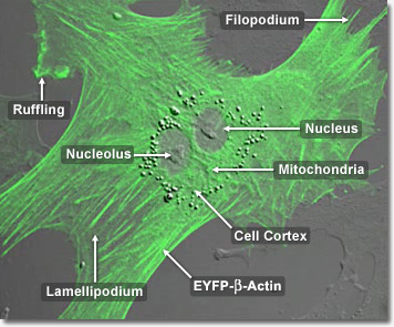
Actin has the capability of quickly assembling into long polymer rods called microfilaments which have a variety of roles. They interact with myosin to permit movement of the cell, form part of the cell’s cytoskeleton, and they pinch the cell into two during cell division. In muscle contraction, filaments of actin and myosin alternately unlink and chemically link in a sliding action. The energy for this reaction is supplied by adenosine triphosphate, ATP. Actin is present in most cells, especially muscle cells and contributes to the cell’s structure and movement.