Anywhere from one to several thousand mitochondria may be present in a cell, depending upon the metabolic requirements of that cell. Mitochondria are found in nearly all eukaryotes, including animals, fungi, plants and protists and are large enough to be observed with a light microscope. The name of the organelles was coined to reflect the way they appeared to the first scientists to observe them, stemming from the Greek words for “thread” and “granule.”
Video 1 - Run Time: 64 Seconds
Video 2 - Run Time: 31 Seconds
For several years after their discovery, mitochondria were commonly believed to transmit hereditary information. It was not until the mid-1950s, when a method for isolating the intact organelles was finally developed, that a firm understanding of the biochemistry and molecular biology surrounding mitochondrial function was established. In the digital video presented above, normal baby hamster kidney fibroblast cells (BHK line) are expressing a fusion of DsRed2 fluorescent protein to a mitochondrial targeting signal peptide.
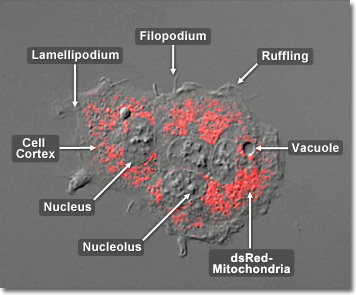
Two highly specialized membranes, each with very specific duties, help mitochondria function. The outer membrane, containing the protein porin, serves as a sieve, filtering out unneeded molecules. The inner membrane is substantially larger and more selective than the outer membrane and contains many folds or cristae that envelop the inner matrix. This membrane allows only selected molecules through and uses transport proteins located along the membrane to regulate and limit which ones are allowed to pass. The number of cristae, or infolds, is directly related to the energy needs of the cell. Since the two membranes work together along with specialized proteins and compartments to form adenosine triphosphate (ATP), more folds on the inner membrane increases the membranal surface area and, therefore, the mitochondrion’s capacity to generate ATP. In the digital video presented above, normal baby hamster kidney fibroblast cells (BHK line) are expressing a fusion of DsRed2 fluorescent protein to a mitochondrial targeting signal peptide.


