In general, fluorescent probes are classified according to their excitation and emission characteristics, as well as their chemical and biological properties. The following tabulation reviews examples of probes in each of the important biological classes.
Proteins, Nucleic Acids, Saccharides, Lipids and Toxins
- Agonists/Antagonists: TRITC-labeled alpha-bungarotoxin - The distribution of receptors can be observed at neuromuscular junctions when alpha-bungarotoxin binds specifically with acetylcholine receptors.
- Antibodies: FITC-labeled anti-mouse IgG (or IgG and IgM) antibodies - A selected antibody (secondary antibody) against IgG or IgM (primary antibody) from various animals is labeled with fluorescent dyes.
- Avidin: Texas Red-labeled streptavidin (or avidin) - This probe can be used to detect the biotin-labeled FISH probe or antibody because the avidin can bind specifically with the biotin.
- Dextran: Texas Red-labeled dextran - Dextran, a polysaccharide with a high molecular weight of 3,000 to 70,000, is labeled with a fluorescent dye or probe (for example, a calcium ion probe, pH probe, or fluorescein). The labeled polysaccharide is then injected into a cell and distributed uniformly in the cytoplasm.
- Lipids: NBD-labeled phosphocholine - Lipids labeled with the fluorescent probes are mixed with the membrane lipid to measure the membrane fluidity by means of photobleaching techniques (primarily recovery after photobleaching, FRAP), and to observe the process of membrane fusion.
- Nucleotides: Cy3 labeled dCTP - Used in labeling of fluorescence in situ hybridization (FISH probes (by means of Nick translation). Nick translation is a technique of labeling DNA fragments for FISH probes. The reagents are usually provided in the form of a kit by the manufacturer. The DNA catabolic enzyme (DNase 1) randomly nicks one of the two DNA strands in the primary reaction, and a second enzyme (DNA polymerase 1) digests the DNA from the nick and simultaneously synthesizes DNA at the digested portion. An expanse of newly synthesized DNA contains fluorescent dye-labeled nucleotides. The biotin nucleotide (biotin-16-dUTP) or digoxigenin nucleotide (dioxigenin-11-dUTP) can be used in place of the fluorescent dye-labeled nucleotide.
- Phallotoxin: Rhodamine phalloidin - Bundles of actin filaments (a stress fiber) can be observed in cells because fluorescently labeled phalloidin and the phallacidin can bind specifically with actin filaments (microfilaments).
Staining of Lipid Membranes
- Membrane Potential-Sensible Probes: WW 781, RH-155, and Di-8-ANEPPS - These dyes are incorporated into the cell membrane. Their absorption or fluorescence intensity varies depending on the membrane potential. Fast-type dyes with response rates on the order of milliseconds should be used.
- Organelle Membrane-Specific Probes: DiOC6(3), NBD ceramide, Rhodamine 123 - These dyes stain specifically the organelle membranes, such as the endoplasmic reticulum membrane, Golgi membrane, and mitochondrial membrane.
- Tracer Probes: DiI, DIO, DIA - These dyes are incorporated into the cell membrane and are used in the observation of nerve extension.
Staining of Cytoplasm
- Ion Concentration-Sensible Dyes: Fura-2, Indo-1, Fluo 3, BCDCF, and SNARF-1 - The membrane-permeable acetoxymethyl ester of the probes is easily introduced into living cells. Their fluorescence intensities and excitation/fluorescence spectra vary depending on the intracellular ion concentration (for example, calcium ions and pH).
- Tracer Dyes: Sulforhodamine 101 and Lucifer Yellow CH - These water-soluble dyes are micro-injected into the cell for the observation of the extension of nerve cells.
- Live Cell Probes: Calcein AM and CFDA (Carboxyfluorescein Diacetate) - Esterases in cells turn the membrane-permeable dyes into membrane-impermeable dyes, thereby trapping them inside the cells.
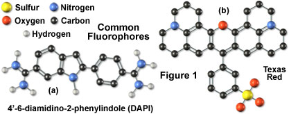
- Staining of Nucleic Acids: DAPI, Propidium iodide, Hoechst 33342, YOYO-1
- These dyes bind with nucleic acids (DNA, RNA) inside the cell and become fluorescent. They are used to observe the morphology of nuclei and chromosomes and to determine DNA quantity.
- Fluorescent Proteins: GFP (Green Fluorescent Protein and derivatives)
- This protein was originally isolated from jellyfish and emits green fluorescence under blue-light excitation. The GFP gene has been isolated and improved to obtain different color variants.
- Fluorescent Enzyme Substrate: Dihydrorhodamine 123
- The activity of an enzyme can be measured on a microscope as the dye becomes fluorescent by the specific enzyme in the cells.
- Fluorescent Latex: FluoSpheres (Molecular Probes)
- Polystyrene beads having a diameter between 0.01 and 15 micrometers are used in the observation of the transport in axons, research into phagocytosis, and measurement of blood flow, or as the standard for determining resolution and microscopy performance in fluorescence microscopy.


