Multiphoton Excitation Laser Scanning Microscopy
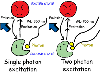
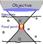
In multiphoton excitation microscopy, fluorescent dyes are excited by absorbing the energy of two or more photons simultaneously (see Figure 1). For two-photon excitation, The dyes are excited at around twice the wavelength used in ordinary fluorescence observations, since the photon energy is inversely proportional to the wavelength. Because the excitation probability of the fluorescent dye in two-photon excitation is proportional to the square of excitation light intensity, only the area proximal to the focal point, where the photon density is high, can be excited; tomographic images can then be obtained by scanning the laser beam (Figure 2, in which the non-excited area is grayed). The multiphoton excitation laser scanning microscopy provides similar tomographic images to the confocal LSM through the different principles.
Using probes such as calcium ion indicators, some of which are excited by ultraviolet light, considerations for the aberration and transmittance of ultraviolet light through the microscope optics can be eliminated in multiphoton excitation microscopy. Specimen damage from the ultraviolet light can also be reduced. Multiphoton excitation is expected strongly to photo-activate caged compounds only at a three-dimensionally specific point in specimens.
Figure 1
| Figure 2
|
Equipment Configuration
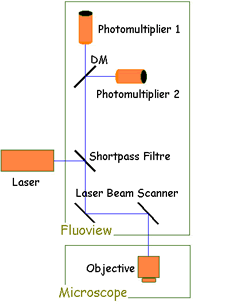
A confocal laser scanning microscope must be modified and equipped with a specialized near-infrared pulsed laser system in order to perform multiphoton imaging. The following characteristics should be used as a guide when comparing multiphoton excitation with conventional laser scanning confocal microscopy.
|  |
Applications of Multiphoton Microscopy
- Applications based on the advantages of avoiding ultraviolet light:
- Calcium ion concentration measurement using Indo-1.
- DNA and nucleus staining with DAPI and Hoechst.
- Applications based on the advantages of using near-infrared light:
- Deep fluorescence observation in thick specimens.
- Applications based on the advantages of local light absorption:
- Photo-activation of caged compounds.
- Fluorescence recovery after photo-bleaching.
Bibliography
- Denk, W., Proc. Natl. Acad. Sci. USA, 91: 6629 (1994).
- Svoboda, K., et al., Science, 272: 716 (1996).
- Sako, Y., et al., Journal of Microscopy, 185: 9-20 (1997).
Internet Resources
Haruo Kasai Laboratory - The Kasai laboratory has developed new approaches, based on two-photon excitation microscopy, to the study of neurons and secretory cells. The application of these approaches has provided important insight into the structure-function relations of central synapses and the mechanisms of exocytosis in both neurons and secretory glands such as pancreatic islets and acini.
Sorry, this page is not
available in your country.


