Products Search
Inverted Microscopes
APX100
- Easy-to-use, all-in-one microscope system
- Publication-quality images in a few clicks
- Fast, efficient data management
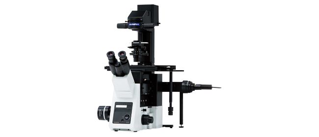
CKX53
The CKX53 microscope eases the cell and tissue culture workflow, simplifying steps such as live cell observation, cell sampling and handling, image capture, and fluorescence observation. Its integrated phase contrast system, compact, ergonomic design, and stable performance enable simple, efficient cell observation. The universal sample holder and expandable stage accommodate a wide variety of cell culture container types and sizes.
- Precentered phase contrast
- Inversion contrast (IVC) technique provides clear three-dimensional views
- Fluorescence with a 3-position slider
- View multilayer tissue flasks up to 190 mm (7.5 in.) in height up thanks to the detachable condenser
CrestOptics Confocal Series
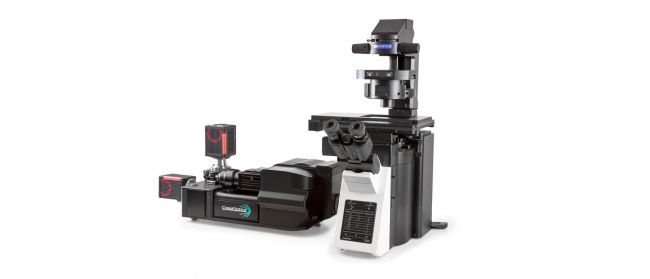 |
CrestOptics solutions are flexible and powerful spinning-disk confocal systems for live-cell imaging. Combined with Evident’s range of microscopes and optics and 89 North’s laser diode illuminators, these customizable systems give researchers a powerful platform to expand their live-cell imaging capabilities.
- CICERO: Improve your widefield microscope imaging capabilities
- X-Light V3: Simultaneous dual camera imaging for challenging live-cell applications
- Flexible laser lines, including NIR, catered to your research needs
IXplore Live
Designed to reduce photobleaching and phototoxicity, the IXplore Live system is optimized for physiological experiments involving live cell and tissue observation. Offering precise environmental control and enhanced rigidity, it supports long-term cell viability and stability for time-lapse imaging applications, such as in cancer, stem cell, and brain research.
- Maintain focus accurately and reliably in time-lapse experiments with TruFocus™ Z-drift compensation system
- Discover the real morphology of your cells with Olympus silicone immersion optics
- Real-time controller helps limit cell disturbance, enabling physiologically relevant data
IXplore Pro
The IXplore Pro automated system acquires panoramic, multichannel images without a complex workflow. Easing experiment setup, the Graphical Experimental Manager offers fully automated multidimensional observation capabilities. The microscope’s high-precision ultrasonic stage, automated focusing, and bright, uniform fluorescence illumination facilitate image stitching applications.
- Automated multidimensional observation (X, Y, Z, T, wavelength, and positions)
- Boost your statistics with multiwell plate screening
- Acquire fluorescence panoramic images of large samples, such as brain slices
- Create 3D optical sections and enhance resolution with TruSight™ deconvolution
IXplore Spin
The IXplore Spin system features a spinning disk confocal unit that enables fast 3D image acquisition, a large field of view, and prolonged cell viability in time-lapse experiments. Researchers can use it to perform rapid 3D confocal imaging with high resolution and contrast at greater depths for imaging into thicker samples. The spinning disk also helps to cut down on photobleaching and phototoxicity of samples upon excitation.
- Real-time controller (U-RTCE) helps optimize the device’s speed and precision during automated acquisition
- TruFocus™ Z-drift compensation system maintains focus for each frame
- Precise 3D imaging with improved light collection using X Line™ objectives
- Upgrade to the IXplore SpinSR super resolution system as your research progresses
IXplore SpinSR
The IXplore SpinSR system is our confocal super resolution microscope optimized for 3D imaging of live cell specimens. Like the IXplore Spin system, it has a spinning disk system for fast 3D imaging while limiting phototoxicity and bleaching. However, it achieves super resolution images down to 120 nm XY and enables you to switch between widefield, confocal, and super resolution with the click of a button.
- Sharp, clear super resolution imaging down to 120 nm XY, owing to Olympus Super Resolution (OSR)
- Prolonged cell viability in confocal time-lapse imaging due to less phototoxicity and bleaching
- Use two cameras simultaneously to achieve fast, two-color super-resolution imaging
- Super resolution imaging with the world’s first plan apochromat objectives with a numerical aperture (NA) of 1.5*
* As of November 2018. According to Olympus research.
IXplore Standard
 |
Optimized for basic multicolor fluorescence imaging and routine experiments, the IXplore Standard system is easy to operate and ergonomically designed. Even with standard cell culture vessels, it captures high-quality, publication-worthy images while providing accurate and repeatable results at high magnifications. The IXplore Standard system’s simplified workflow and ease of use facilitate a wide range of standard imaging applications.
- High repeatability and accuracy for standard imaging tasks
- Benefit from the same optical capabilities found in high-end IXplore systems
- Easily upgrade to encoded functionality to boost the reproducibility of experiments
IXplore TIRF
For membrane dynamics, single-molecule detection, and colocalization experiments, the IXplore TIRF system enables sensitive simultaneous multicolor TIRF (total internal reflection fluorescence) imaging for up to four colors. Olympus’ cellTIRF system provides stable motorized individual laser-angle control, providing equal evanescent wave penetration for high-contrast, low noise images. Our TIRF objectives feature high SNR, high NA, and correction collars to adjust for cover glass thicknesses and temperature.
- Exact colocalization of up to four markers thanks to individual penetration depth control
- Take advantage of Olympus’ TIRF objective with the world's highest NA of 1.7*
- Intuitive setup of complex experiments with the Graphical Experiment Manager (GEM), cellFRAP, and U-RTCE
* As of July 25, 2017. According to Olympus research.
Sorry, this page is not
available in your country.