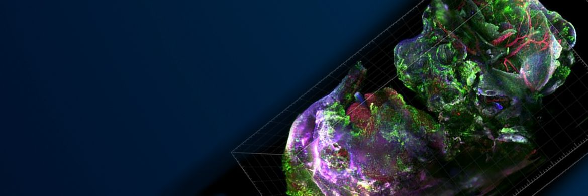
Imaging Optically Cleared Samples
We live in a three-dimensional world. However, observing our world using conventional microscopy techniques can be challenging since most biological models beyond a few hundred microns in thickness become optically opaque due to destructive light scattering.
To overcome this limitation in the past, researchers needed to laboriously section and digitally reconstruct these samples. Optical clearing has become a necessary tool for 3D imaging since these techniques homogenize the scattering of light within tissue samples, enabling us to create more accurate 3D tissue models.
But with many different strategies for imaging optically cleared samples—from confocal, multi-photon, to light sheet microscopy—it can be difficult to determine which imaging solution is the best match for your needs.
In the first part of our webinar series, we will discuss optical clearing of biological samples and its benefits. We will also explore how to image these samples using various microscopy techniques and the advantages and disadvantages of each method to help you determine which system best suits your needs.
Agenda:
- What is optical clearing, and what are the benefits?
- Tips and tricks for sample clearing
- Imaging cleared samples: matching equipment to your imaging needs
- Conclusions
Presenters:
 | Tim Steppe, PhD |
 | Gulpreet Kaur, PhD |
Sorry, this page is not
available in your country.
