MetaMorph for Olympus Overview
Proven Performance for Image Capture Display and Analysis
MetaMorph for Olympus converts your biological experiment into a publishable result. Built on the market-proven MetaMorph platform, this powerful package offers functionality, flexibility, and stability that meet the most demanding image acquisition and analysis needs. With over 10,000 publications citing MetaMorph, you can rest assured that your imaging needs will be met now and in the future.
Acquisition & Device Control
MetaMorph for Olympus device control makes acquisition fast and easy, allowing the integration of IX2 and BX3 microscopes and a variety of other hardware devices into a single, intuitive acquisition interface.
Image Display & Processing
Image display and processing tools are crucial for performing accurate image analysis, and MetaMorph for Olympus offers a complete toolbox of advanced features in this area, from background subtraction and shading correction, to morphology filters and an interactive 4D Viewer.
Image Analysis
MetaMorph for Olympus offers analysis tools to handle everything from simple intensity logging to advanced morphometry analysis, colocalization, FRET, 3D measurements, and more. Optional Application Modules perform assay-specific analysis, such as counting nuclei or assessing cell cycle phases, in a simple wizard-like interface.
Customization
Powerful and easy-to-use journal (macro) functions in MetaMorph for Olympus enable users to further automate acquisition, processing, and common analysis routines, while custom toolbars and menus provide users quick access to commonly used menu options, taskbars, and journals.
MetaMorph for Olympus Packages
The MetaMorph for Olympus is available for both acquisition and offline (analysis only) needs.
MetaMorph for Olympus Acquisition Features | Drivers for many popular CCD cameras, Olympus IX2 and BX3 motorized microscopes plus shutters, filterwheels, and XY stages from various manufacturers Multi-dimensional acquisition interface |
|---|---|
MetaMorph for Olympus Acquisition Options | Simultaneous and split camera acquisition Automated slide scanning and stitching Multi-well plate interface Control of various devices for FRAP, uncaging, photobleaching and photoablation devices |
MetaMorph for Olympus Acquisition and Offline Features | Multi-dimensional data display interface Montage display Image processing features such as shading correction and kernel filters Integrated Morphometry Analysis for detection and analysis of objects Colocalization and FRET tools |
MetaMorph for Olympus Acquisition and Offline Options | 4D viewer Motion tracking Multi-well data viewing Application Modules for specialized application analysis needs |
Additionally, we offer MetaFluor for Olympus for ratio imaging of intracellular ion concentrations.
For product and sales information, please contact: metasales@olympus.com
Application Modules
MetaMorph for Olympus offers eleven user-friendly Application Modules for biology-specific analysis. Each Application Module features a dedicated dialog box with intuitive setting selections, improved segmentation through adaptation to local content, and both field and cell-by-cell data logging. After analysis is complete, users can interactively view tabular and image results side by side. Application Modules may be incorporated into macros for increased customization and automation of analysis. All Application Modules are validated in-house and with third-party collaborators.
Angiogenesis Tube Formation
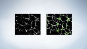
- Better quantitation by creating a single in-focus composite image from multiple Z-series images
- Multi-parameter analysis measurements include tube length, number of branch points, number of nodes and more
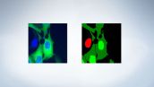
Cell Cycle
- Classification and quantification of cells in different stages of the cell cycle
- Option to use specific apoptosis and mitotics stains, for increased classification accuracy
Cell Health
- Analysis of up to three fluorescent probes for cell-based apoptosis and necrosis assays
- Classification and quantification of viable, early and late apoptotic cells, necrotic cells
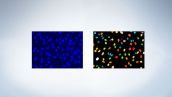
Cell Scoring
- Identification of two sub-population of cells
- Ideal for counting and logging measurements of cells in two-wavelength experiments
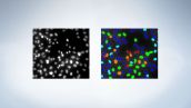
Count Nuclei
- Automatically counts nuclei and captures intensity measurements
- Accurate segmentation of touching cells
Granularity
- Count punctuate objects
- Designed for receptor internalization or clustering target molecules
- Choice of six granularity indices
- Measurements include count, total and mean area, and intensities of granules and nuclei
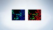
Live/Dead
- Classification and quantification of live and dead cells
- Measurements include counts and percentages of live and dead cells, per-wavelength measurements and more
Mitotic Index
- Classification and quantification of mitotic and interphase cells
- Measurements include count, percentage and wavelength-specific intensities of mitotic and non-mitotic nuclei, and more
Monopole Detection
- Classification and quantification of mitotic cells with monopolar or bipolar spindles
- Measurements include count and percentage of monopoles, bipoles and interphase cells, DNA structures area, cell classification and more
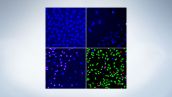
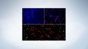
Multi Wavelength Cell Scoring
- Multi-parametric analysis of up to seven wavelengths
- Identification of sub-populations of cells
- Measurements include scoring profiles, wavelength-specific count and percentage of negative and positive cells, cell-by-cell wavelength-specific stained area, integrated and average intensities.
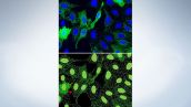
Neurite Outgrowth
- Designed for the measurement and analysis of neurite outgrowths
- Works with or without nuclear stain
- Measurements include total neurite outgrowth, total branches and cell bodies, straightness and more
Kinase activation (Cell Scoring)
Fatty acid uptake (Cell Scoring, Multi Wavelength Cell Scoring)
Mitosis (Mitotic Index, Cell Cycle)
Adipogenesis (Cell Scoring, Multi Wavelength Cell Scoring)
Examining transfection efficiencies (Cell Scoring, Multi Wavelength Cell Scoring)
Studying intracellular structures (Granularity)
Receptor internalization (Granularity)
Clustering target molecules(Granularity)
Process extension (Neurite Outgrowth)
Neurodegenerative or neuroregenerative diseases (Neurite Outgrowth)
Cell differentiation (Stem cell research) (Neurite Outgrowth)
Protein expression & modification (Cell Scoring, Multi Wavelength Cell Scoring)
Transient transfection (Cell Scoring, Multi Wavelength Cell Scoring)
Detect monopolar spindles (Monopole Detection)
Cell signaling (Cell Health, Cell Cycle)
MetaFluor for Olympus
Fluorescence ratio imaging is the monitoring of live cells in which a fluorescent indicator of intracellular ions is introduced. Indicator dyes have been designed to shift their fluorescence excitation or emission spectrum when binding with specific ions. Images are obtained at two different wavelengths, typically matching the absorption bands at the high and low binding conditions.
By rationing the intensities in the images, it is possible to construct a map showing the local ion concentrations throughout the field of view. Since the monitoring process is nondestructive, image acquisition can be repeated frequently to trace and monitor the time course of cellular responses.
MetaFluor for Olympus is designed for dual-wavelength intracellular ion measurements. The system provides simultaneous display of the raw data, ratio image, graphs of intensities, ratios and ion concentrations, and a non-ratiometric image such as a brightfield or phase-contrast image. Two different ratiometric indicators can be imaged and measured simultaneously.
Custom Configuration
Toolbars, menus, wizards and dialog boxes help move you through the image processing steps quickly. Features such as multiple image windows, flexible device control, synchronization and timing, and journals allow for automated image acquisition and analysis unlike any other system.
With MetaFluor for Olympus, you customize the set-up once, then let the experiment run by itself. You are able to collect a large amount of data online and process it with either MetaFluor for Olympus or an analysis-only copy of the software.
Monitoring of Live Cells
Regions of interest can be generated automatically or manually placed on your image to monitor intensity, ratio value or ion concentration. Measurements are then made simultaneously on all the regions of interest and update continuously on a scrolling graph, allowing you to follow dynamic changes as they occur in your living samples.
A display of multiple graphs gives flexibility in the presentation of your experiment's data. MetaFluor enables you to click on graph traces to display a readout of the time and data value for the region nearest to the click.
The Event Mark function is useful to record when drugs or solutions are added, experimental conditions changed, triggers are received or sent or other events occur. You have the option to associate a timer and an alarm bell to each event. Additionally, for perfused samples, ambient conditions can be logged and tracked. Each image has an annotation that is saved within the TIFF file format. The annotation will record wavelength-dependent settings. Additional information can be stored in a protocol file.
Export Data for Analysis
If needed, MetaFluor for Olympus can log and export all measurements to either a text file or to a spreadsheet program such as Microsoft Excel.
Compatible with MetaMorph for Olympus
Because MetaFluor for Olympus saves images in TIFF file format, you can import them into MetaMorph for Olympus for further processing and analysis.
Presentation and Publication
Images in MetaFluor for Olympus can be displayed in monochrome, pseudocolor, or using a variety of user-defined set of values. Ratio images can also be displayed using a special display mode called Intensity Modulated Display, or IMD. With the IMD mode, color is used to represent the relative ratio value, while the intensity or brightness of the color is used to represent whether the brightness of the source images. This technique helps automate the process of extracting spatial information from the background, by automatically eliminating background fluorescence from the scene. MetaMorph and MetaFluor are trademarks of Molecular Devices, Inc.