Not Available in Your Country
Sorry, this page is not
available in your country.
Overview
 | Super Resolution with Nanometer PrecisionOlympus and Abbelight have joined forces to provide researchers with advanced and intuitive nanoscopy imaging systems. Abbelight’s vast expertise in single-molecule localization microscopy (SMLM) and Olympus’ rich history in optical precision form the foundation of this collaboration. By combining Abbelight’s SAFe nanoscopes with our IX™ series microscopes, users can transform their Olympus inverted microscopes into multimodal powerhouses that feature both SMLM and total internal reflection fluorescence microscopy (TIRFM) in one system. Get in Touch |
|---|
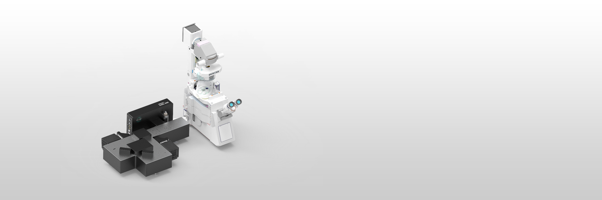
Modular Nanoscopy SolutionsThe SAFe nanoscopes can be incorporated onto any Olympus inverted microscope that features a camera port, making it an easy and flexible nanoscopy solution. Our IX83 microscope is well suited for nanoscopy applications due to its stable frame, TruFocus™ Z-drift compensation module, and open structure. SAFe nanoscopes can also be combined with our FV3000 confocal laser scanning microscope and the IXplore™ SpinSR super-resolution microscope system, enabling researchers to maximize their imaging capabilities with confocal microscopy, TIRFM, and SMLM in one system. | 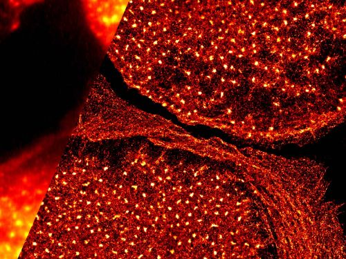 |
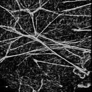
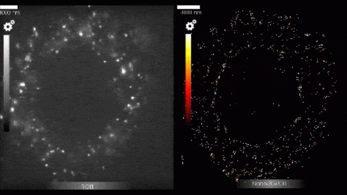 | What is SMLM?Single Molecule Localization Microscopy (SMLM) uses various techniques to cause individual fluorescent molecules to “blink.” These individual blinks are processed to produce precise, high-resolution images (down to 10nm) showing the 3D coordinates of single molecules. Using SMLM, users can access new avenues for both spatial and temporal analyses at the nanometer scale. |
A Complete SMLM SolutionAbbelight provides a complete solution enabling even SMLM novices to achieve successful results during their first experiment. Preparation: A good experiment starts with good sample preparation. Abbelight offers ready-to-use and optimized SMLM kits for dSTORM and photo-activated localization microscopy (PALM). Imaging: Each system can be customized and upgraded to meet user needs and is run on Abbelight’s intuitive and easy-to-use software. Analysis: Abbelight’s NEO software offers a complete analysis workflow helping users to translate their data into meaningful results. Support: Throughout the experimental workflow, a dedicated Abbelight expert is assigned to support each user. Users can also acquire expertise through the online Abbelight Academy, which offers guides, video tutorials, and best practices. |
|
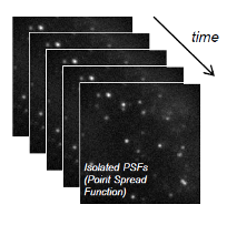
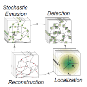
How Single-Molecule Localization Microscopy WorksSMLM relies on the ability to stochastically activate fluorescent molecules to distinguish them spatially. Consecutive images are acquired as different molecules fluoresce, and the accumulated raw data are processed in real time to localize every single molecule with nanometer precision (down to 10 nm). Abblelight systems work with a range of techniques including dSTORM, PALM, and PAINT, to enable SMLM with both live and fixed cells, and a wide range of commonly used fluorophores. These techniques only differ in how the fluorophore activation/inactivation is induced. For users new to SMLM, Abbelight’s experts will guide you through developing a protocol specific to your application.
|
Need assistance? |
Applied Technologies
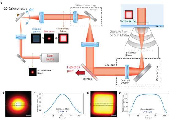
ASTER Technology for High Localization Precision over a Wide Field of ViewThe SAFe nanoscopes all share the same unique excitation system, which is based on the Adaptable Scanning for Tunable Excitation Regions (ASTER) technology.1 ASTER generates homogenous illumination in TIRF, HiLo, and EPI modes while performing SMLM modalities such as PALM, STORM, or PAINT with a localization precision reaching 10–15 nm in 3D on a field of view (FOV) of 150 x 150 µm2. |
Schematic of ASTER and the resulting illumation patterns. | The ASTER illumination method offers a novel capability to exploit the entire FOV of sCMOS cameras for SMLM and TIRF imaging. ASTER uses two galvanometer mirrors to control illumination at the sample plane. While the excitation beam keeps its position in the back focal plane (BFP), an angular rotation of a galvanometer induces a similar angle in the BFP, corresponding to a different position in the sample plane. By applying specific patterns, such as raster scanning, ASTER can provide uniform excitation on tunable FOVs of up to 150 × 150 µm2 for all excitation modes (EPI, HiLo, and TIRF). |
Wide FOV TIRF Imaging with Homogenous IlluminationOlympus is a pioneer in the TIRF microscopy field, and our range of TIRF objectives is designed to provide tight control over the evanescent wave produced in TIRF imaging with magnifications ranging from 60X to 150X. The APON100XHOTIRF objective has the world’s highest NA of 1.7*, while the UPLAPO60XOHR and UPLAPO100XOHR are the world’s first plan apochromat objectives with a NA of 1.5.* With Olympus’ optics and Abbelight’s ASTER illumination technology, users can achieve homogenous TIRF illumination over a wide FOV. *As of November 2018. According to Olympus research. | 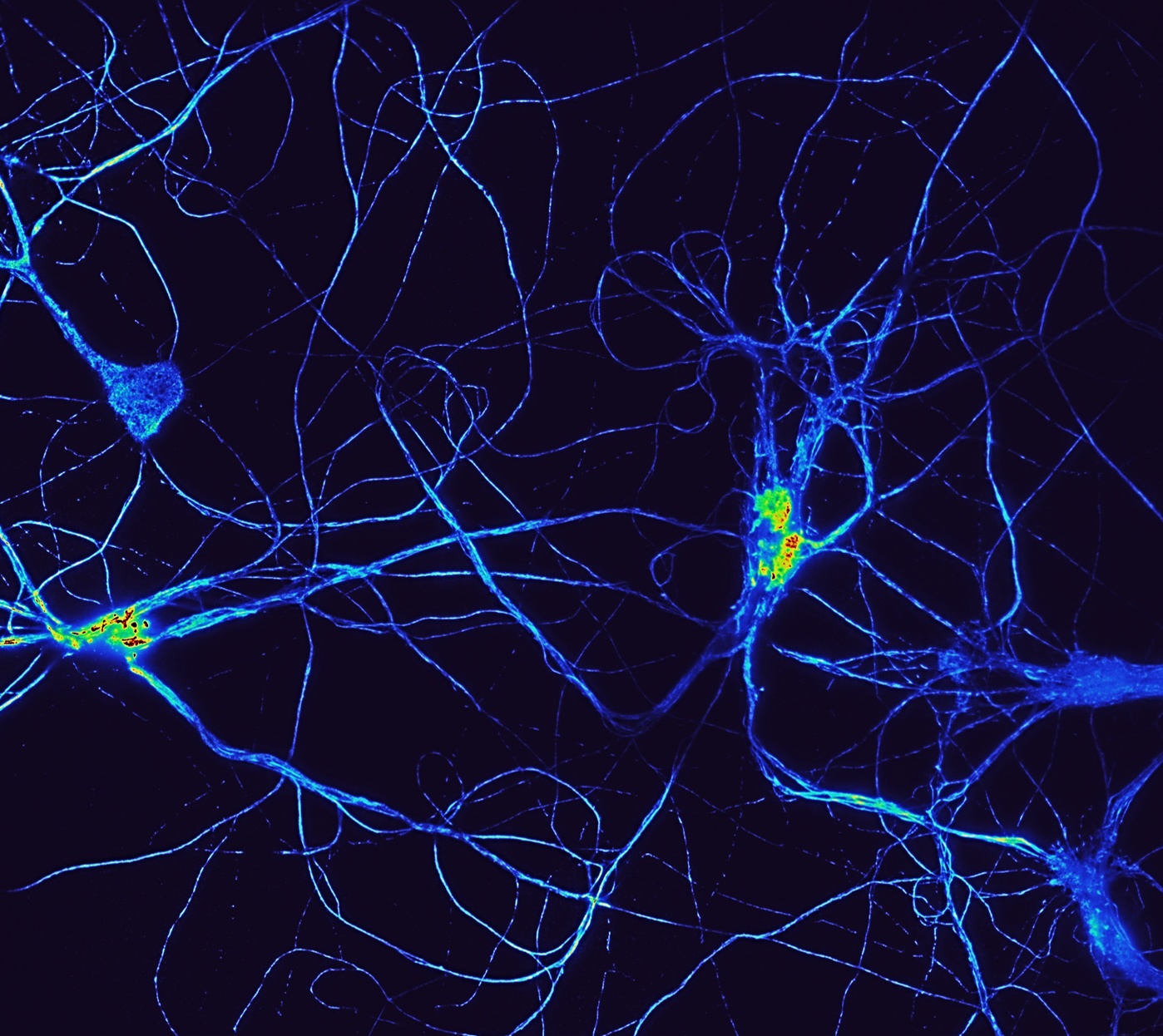 Cultured hippocampal neurons stained for spectrin cytoskeleton and imaged in TIRF microscopy mode. Homogeneous TIRF over the entire field of view of a Hamamatsu Fusion sCMOS camera (larger than the camera port size) was achieved using Abbelight ASTER technology. Sample courtesy of C. Leterrier, NeuroCytoLab, Marseille, and images by Adrien Mau, ISMO, Orsay. |
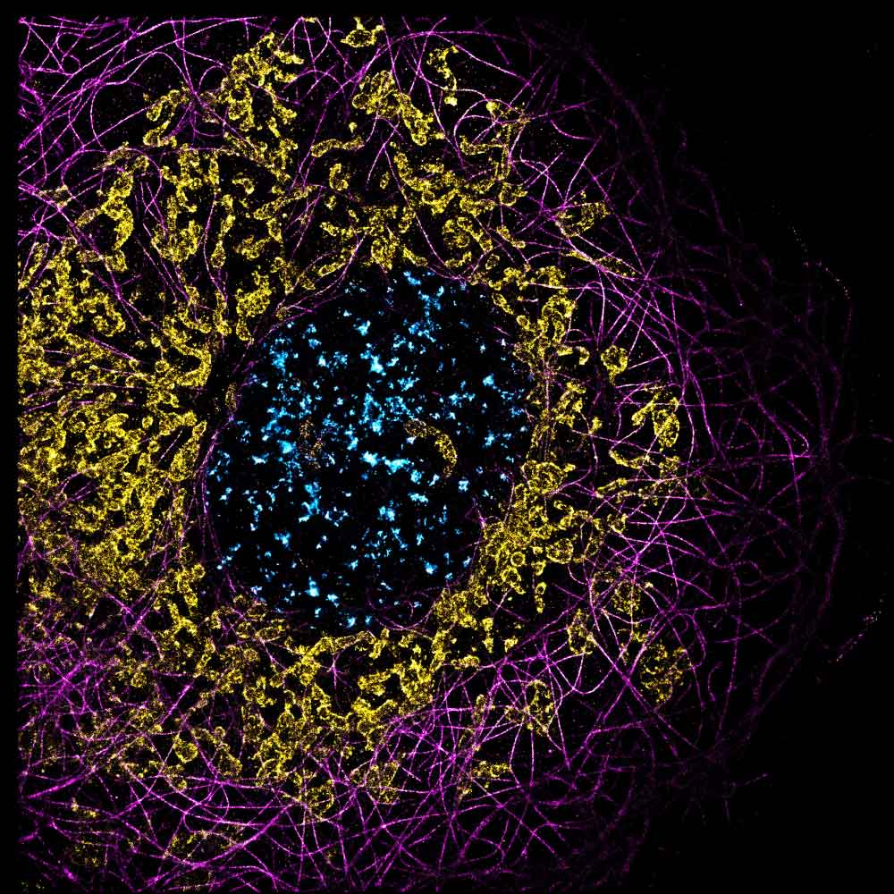 U2OS cells stained for microtubules (alpha-Tubulin antibody) CF660, mitochondria (anti-TOMM20) CF680, and chromatin (EdU) AF647. Simultaneous multicolor 2D dSTORM with spectral demixing. | Spectral Demixing: Multicolor Imaging with One Laser, One Buffer, and One AcquisitionAlthough 3D nanoscopy has revolutionized the fluorescence microscopy field by attaining unprecedented resolutions, multicolor imaging remains challenging in SMLM. This difficulty is due to several factors, including chromatic aberrations, the choice of buffers, and the choice of single-molecule-compatible dyes. To solve this challenge, Abbelight has implemented spectral demixing for SMLM. By separating far-red dyes using a dichroic cube and ratiometric algorithms, spectral demixing elegantly enables simultaneous multicolor imaging in SMLM. |
References1: A. Mau, K. Friedl, C. Leterrier, N. Bourg, and S. Lévêque-Fort. Fast scanned widefield scheme provides tunable and uniform illumination for optimized SMLM on large fields of view. Nature Communication. May 21, 2020. |
Need assistance? |
Software
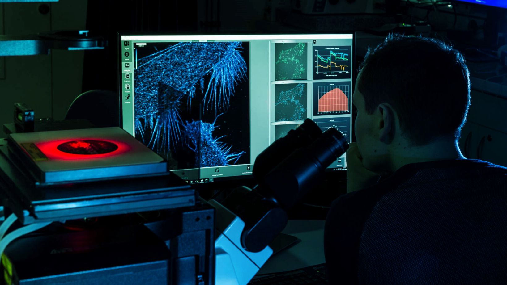
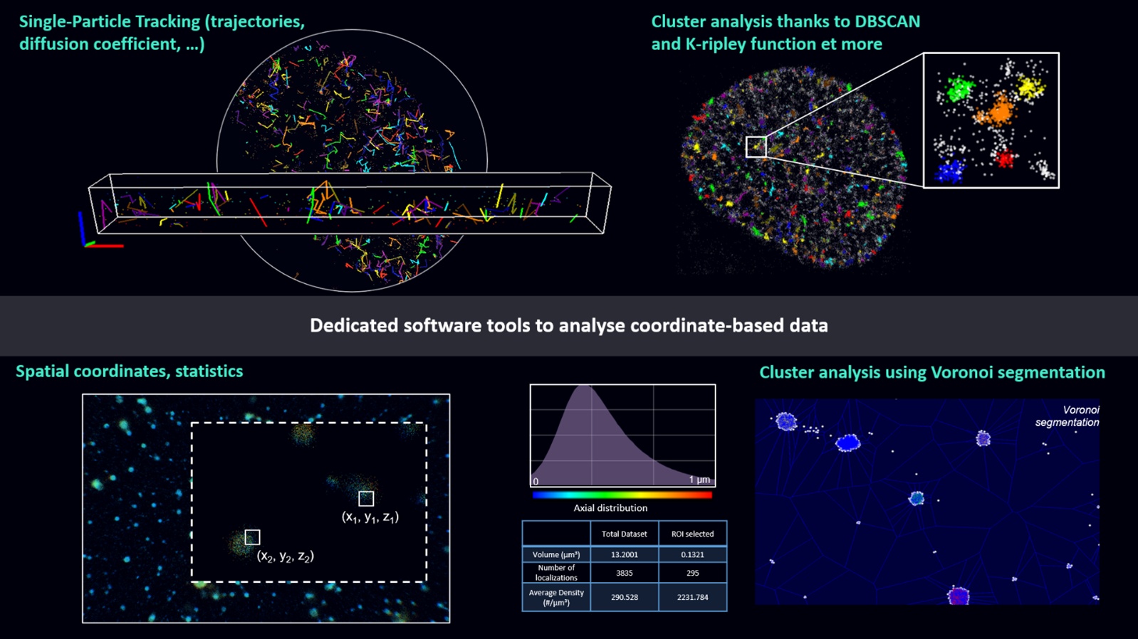
Powerful Software for SMLM Data AnalysisUnlike standard fluorescence microscopy, which generates pixel-based images, SMLM produces point clouds with millions of localizations and associated uncertainties. Abbelight’s NEO software translates this data into a user-friendly package, simplifying data acquisition, and providing real-time image reconstruction and quantitative feedback. NEO software also offers powerful tools to process SMLM data to study spatial and temporal distribution of localized single molecules. This includes cluster analysis using DBSCAN, Voronoi, and colocalization algorithms as CBC or single-particle tracking algorithms.
|
Need assistance? |
Configurations
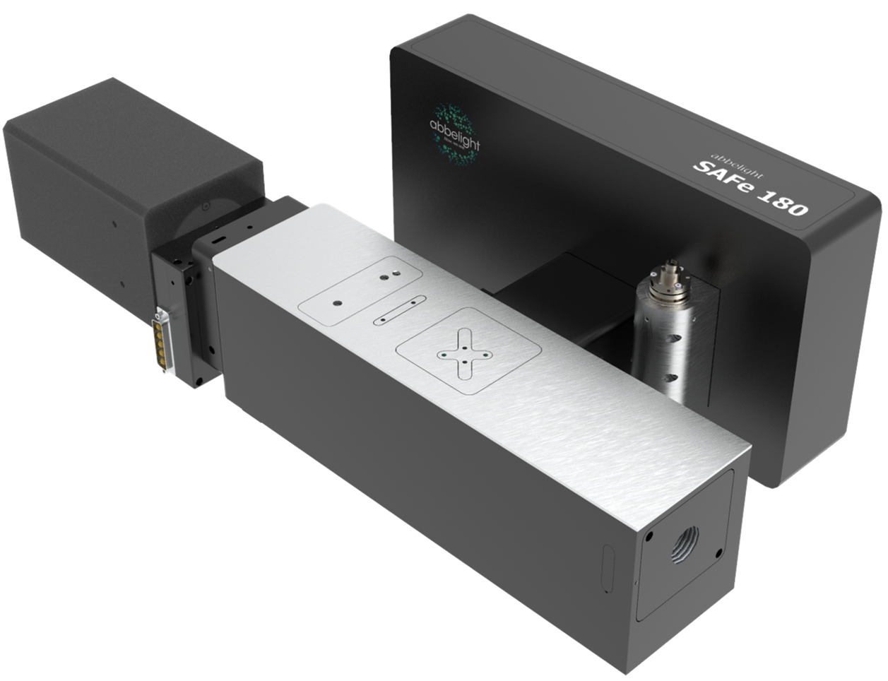
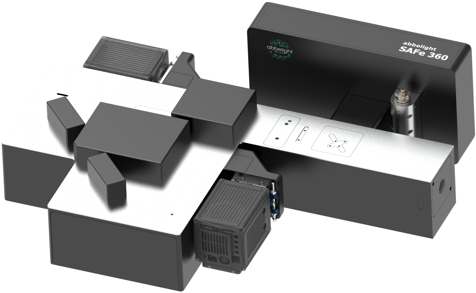
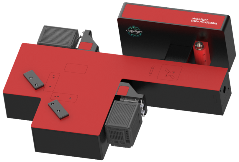
Meet the SAFe Nanoscopes*The three Abbelight nanoscopes include the same SAFe Light illumination module, which integrates ASTER technology for homogenous illumination over a 150 × 150 μm field of view and enables EPI, HiLo, or TIRF excitation modality. *Model availability may vary by region. | 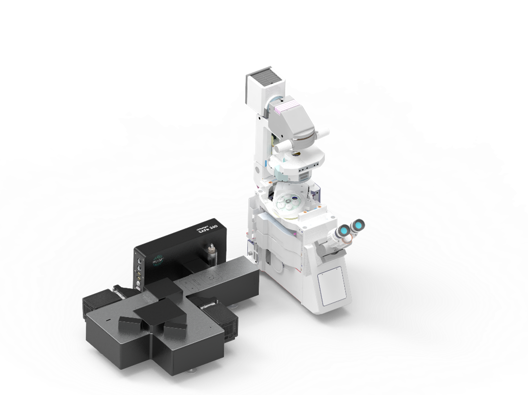 |
| SAFe 180
|
SAFe 360
|
|
| SAFe RedSTORM
|
Need assistance? |
Specifications
Available Systems
|
System Compatibility
|
