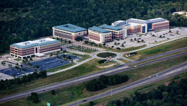
Texas A&M University (TAMU) is a Tier 1 research institution and member of the Association of American Universities, a group of 66 North American research-intensive institutions known for their outstanding credentials in research and education programs. TAMU is the largest university in Texas and the second largest in the US, with an annual enrollment that exceeds 70,000 students, and annual research expenditures in 2020 that exceeded $1 billion. TAMU was among the first four universities in the US to carry the triple designation of a land-, sea-, and space-grant institution, and is currently part of only 17 such universities.
Among the 16 TAMU component colleges, the colleges of liberal arts, sciences, agriculture and life sciences, engineering, and veterinary medicine, as well as the colleges of the Health Science Center (medicine, nursing, public health, and pharmacy) are located near each other on a 5,200-acre campus. The research environment is far-ranging and diverse, with substantial infrastructure investments in engineering, life sciences, and agriculture. TAMU provides a large, vibrant research atmosphere that supports a range of biomedical research relevant to human and animal health.
Innovations that develop from the integration of health care and engineering will increase Texas A&M's ability to personalize care and introduce new options for how care is delivered, extending the reach and impact physicians have on patients. From telemedicine to remote monitoring, these technologies will improve the way patients are diagnosed and treated, and ultimately help make great strides in those patient populations that have been underserved, such as rural area communities and military service members.
Research in the College of Medicine at TAMU aims to improve human life. Using a collaborative mindset and innovative technologies applied to both animal and human model systems, researchers constantly expand their knowledge of how cells, tissues, and organisms function, both when healthy and when compromised by intrinsic genetic defects or external exposures. The college focuses on finding new strategies to treat, alleviate, and prevent human diseases.
Leveraging the unique expertise across both Texas A&M Health and the greater Texas A&M community, the possibilities for multidisciplinary research projects are endless.
This important research is supported through the high-end imaging services of the Integrated Microscopy and Imaging Laboratory (IMIL). The IMIL is the microscopy core facility on the Texas A&M Health Science Center campus. It is located in the newly constructed Medical Research Building II, a 4-story building with over 47,600 sq. ft. of research space. IMIL occupies 2,643 sq. ft. of space dedicated to an optimized microscopy environment.
The IMIL includes three support rooms for sample preparation, cutting-edge microscope systems, and technical support to ensure researchers can successfully incorporate advanced imaging modalities into their existing research programs.
The mission of the IMIL is to support research progress and grant development by encouraging researchers to explore advanced imaging modalities and incorporate them into their existing research programs. The IMIL provides technical expertise and cutting-edge microscope systems to support the research of faculty and staff at the Texas A&M Health Science Center, as well as the teaching mission of the College of Medicine.
Research in the imaging facility includes:
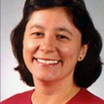
Andreea Trache, PhD
Director, Associate Professor of Medical Physiology and Biomedical Engineering
Dr. Trache’s research focuses on the study of vascular cell adaptation to mechano-chemical stresses in aging and vascular disease. To conduct these studies, she developed microscopy technologies that enable single-cell imaging combined with mechanical stimulation in real time.
Malea Murphy, PhD
Research Specialist II, Technical Staff of the IMIL
Dr. Murphy has diverse developmental biology and stem cell scientific training and extensive experience using microscopy techniques to answer biological questions. She has more than 15 years’ experience with classic sample processing techniques such as cell culture, paraffin and cryosectioning, histological stains, immunological staining of fluorescence and colorimetric assays, as well as imaging of model organisms. She also has extensive experience with cleared tissue imaging and analysis of large tri-dimensional data sets.
Sorry, this page is not
available in your country.