Evident LogoOlympus Logo

Pregunte a los expertos

The Use of Multiplexing in Microscopy for Better Understanding the Skin Immune System in the Context of the Tissue

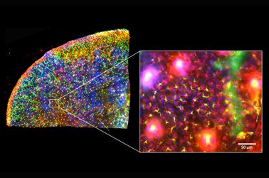
The skin is the first line of defense and the immune system’s biggest barrier for combating pathogens. Being able to accurately characterize and identify immune cell subtypes, tissues structures, and cell distribution in the skin under steady-state conditions provides a powerful tool for understanding the first immunological strategies and biological processes that occur in the presence of pathogens. In this webinar we will review technical aspects involved in the experimental process and explore how complementary imaging technologies might assist us to better understand the immune system.

The presentation is divided into three parts. First, an introduction of the Hugh Green Cytometry Centre will be presented and an overview of the histology and bioimaging technological platforms available. Second, the multiplexing methodology will be discussed, where several topics need to be considered for the design and development of a successful polychromatic panel for microscopy. Finally, preliminary results from a research project will be presented that constitutes part of a diploma program from The Royal Microscopical Society. The project focuses on the identification of immune cell types in the whole mount skin in relation to tissues structures (e.g., blood vessels and lymphatic network). It also centers on the immune cells’ distribution in the tissue as a first barrier of defense against pathogens.

Presenter: Alfonso J. Schmidt
Senior Staff Scientist, Malaghan Institute of Medical Research

Alfonso has a decade of experience working in a Shared Resource Lab (SRL) with a vast knowledge in histology, fluorescent microscopy, and image analysis. His work has been focused in maximizing the capabilities of the equipment available and in creating technical protocols and training modules for the scientific community. Currently, Alfonso oversees the Histology and Bioimaging Facility as part of the Hugh Green Cytometry Centre (HGCC) at the Malaghan Institute of Medical Research. Wellington, New Zealand.  

Productos relacionados

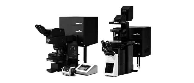
Microscopio confocal de escaneo láser

FV3000

  • Disponible únicamente con las configuraciones de escaneo galvanométrico (microscopio de escaneo FV3000) o híbrida galvanométrica-resonante (FV3000RS).
  • Nueva detección altamente eficaz y precisa en todos los canales mediante la tecnología TruSpectral
  • Optimizada para el tratamiento de imágenes de células vivas proporcionando alta sensibilidad y baja fototoxicidad
  • Inverted and upright frame options to suit a variety of applications and sample types
Escáner de portaobjetos para la investigación

VS200

El escáner de portaobjetos dedicado a la investigación VS200 mejora la velocidad y la eficiencia en aplicaciones que requieren análisis cuantitativos a partir de un alto volumen de muestras, tal y como se experimenta en la investigación del cerebro, el cáncer, las células madre y el descubrimiento de fármacos/medicamentos. Capture con rapidez y facilidad imágenes de portaobjetos de alta resolución y calidad; asimismo, analice, comparta y almacene sus datos con fluidez. El sistema VS200 otorga cinco modos de procesamiento de imágenes: campo claro, fluorescencia, campo oscuro, contraste de fase y polarización simple. Soporte de carga al instante de hasta 210 portaobjetos para escaneos automatizados

  • Procesamiento de portaobjetos completos de alta resolución: de 2X a 100X
  • Admisión de múltiples tamaños de portaobjetos y métodos de observación
  • Proceso de trabajo simplificado y potente: desde el campo claro hasta la fluorescencia multiplexada
  • Exclusiva capacidad de configuración sobre la marcha en el software como en el hardware: desde el escaneo manual de campo claro hasta el reconocimiento por inteligencia artificial (IA), y el escaneo completamente automatizado

The Use of Multiplexing in Microscopy for Better Understanding the Skin Immune System in the Context of the Tissue

The skin is the first line of defense and the immune system’s biggest barrier for combating pathogens. Being able to accurately characterize and identify immune cell subtypes, tissues structures, and cell distribution in the skin under steady-state conditions provides a powerful tool for understanding the first immunological strategies and biological processes that occur in the presence of pathogens. In this webinar we will review technical aspects involved in the experimental process and explore how complementary imaging technologies might assist us to better understand the immune system.

The presentation is divided into three parts. First, an introduction of the Hugh Green Cytometry Centre will be presented and an overview of the histology and bioimaging technological platforms available. Second, the multiplexing methodology will be discussed, where several topics need to be considered for the design and development of a successful polychromatic panel for microscopy. Finally, preliminary results from a research project will be presented that constitutes part of a diploma program from The Royal Microscopical Society. The project focuses on the identification of immune cell types in the whole mount skin in relation to tissues structures (e.g., blood vessels and lymphatic network). It also centers on the immune cells’ distribution in the tissue as a first barrier of defense against pathogens.

Presenter: Alfonso J. Schmidt
Senior Staff Scientist, Malaghan Institute of Medical Research

Alfonso has a decade of experience working in a Shared Resource Lab (SRL) with a vast knowledge in histology, fluorescent microscopy, and image analysis. His work has been focused in maximizing the capabilities of the equipment available and in creating technical protocols and training modules for the scientific community. Currently, Alfonso oversees the Histology and Bioimaging Facility as part of the Hugh Green Cytometry Centre (HGCC) at the Malaghan Institute of Medical Research. Wellington, New Zealand.  

Productos relacionados

Microscopio confocal de escaneo láser

FV3000

  • Disponible únicamente con las configuraciones de escaneo galvanométrico (microscopio de escaneo FV3000) o híbrida galvanométrica-resonante (FV3000RS).
  • Nueva detección altamente eficaz y precisa en todos los canales mediante la tecnología TruSpectral
  • Optimizada para el tratamiento de imágenes de células vivas proporcionando alta sensibilidad y baja fototoxicidad
  • Inverted and upright frame options to suit a variety of applications and sample types
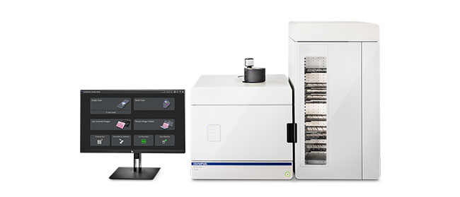
Escáner de portaobjetos para la investigación

VS200

El escáner de portaobjetos dedicado a la investigación VS200 mejora la velocidad y la eficiencia en aplicaciones que requieren análisis cuantitativos a partir de un alto volumen de muestras, tal y como se experimenta en la investigación del cerebro, el cáncer, las células madre y el descubrimiento de fármacos/medicamentos. Capture con rapidez y facilidad imágenes de portaobjetos de alta resolución y calidad; asimismo, analice, comparta y almacene sus datos con fluidez. El sistema VS200 otorga cinco modos de procesamiento de imágenes: campo claro, fluorescencia, campo oscuro, contraste de fase y polarización simple. Soporte de carga al instante de hasta 210 portaobjetos para escaneos automatizados

  • Procesamiento de portaobjetos completos de alta resolución: de 2X a 100X
  • Admisión de múltiples tamaños de portaobjetos y métodos de observación
  • Proceso de trabajo simplificado y potente: desde el campo claro hasta la fluorescencia multiplexada
  • Exclusiva capacidad de configuración sobre la marcha en el software como en el hardware: desde el escaneo manual de campo claro hasta el reconocimiento por inteligencia artificial (IA), y el escaneo completamente automatizado
Expertos
Alfonso J. Schmidt
Senior Staff Scientist
Malaghan Institute of Medical Research

Alfonso has a decade of experience working in a shared resource lab (SRL) with a vast knowledge in histology, fluorescent microscopy, and image analysis. His work has been focused in maximizing the capabilities of the equipment available and in creating technical protocols and training modules for the scientific community. Currently, Alfonso oversees the Histology and Bioimaging Facility as part of the Hugh Green Cytometry Centre (HGCC) at the Malaghan Institute of Medical Research in Wellington, New Zealand.

The Use of Multiplexing in Microscopy for Better Understanding the Skin Immune System in the Context of the Tissueabr 23 2024
Lo sentimos, la página solicitada no se encuentra disponible en su país.
Ask the Expert Sign-up

By clicking subscribe you are agreeing to our privacy policy which can be found here.

Sorry, this page is not
available in your country.

Lo sentimos, la página solicitada no se encuentra disponible en su país.