Nombre: Descripción
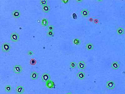
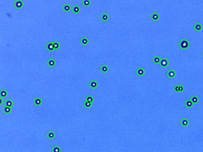
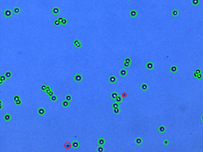
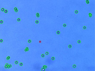
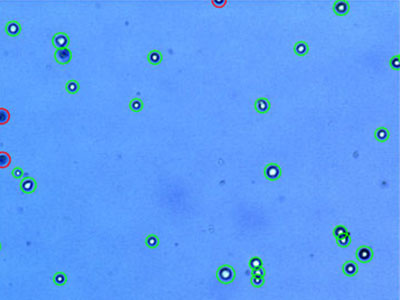
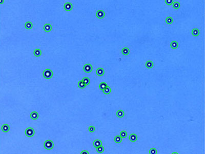
FF hiPS: Células pluripotentes inducidas [iPS] humanas libres de alimentador (línea celular 201B7)
![FF hiPS: Células pluripotentes inducidas [iPS] humanas libres de alimentador (línea celular 201B7)](https://static3.olympus-lifescience.com/data/Image/Product-Gallery/Cell_Counter_R1/product_cell-counter-model_r1_protocol_01.jpg?rev=764D) | |
Protocolo
|
Resultados
|
|---|
Reducción de ruido: 6
Redondez: 60 %
Tamaño celular mín. (μm): 3
Tamaño celular máx. (μm): 60
Nivel de desagrupamiento: Medio
|
Concentración de células totales (células/ml): 1,50 x 10⁶
Concentración de células vivas (células/ml): 1,38 x 10⁶
Concentración de células muertas (células/ml): 1,19 x 10⁵
Viabilidad (%): 92,0
Tamaño celular promedio (μm): 15,9
|
|
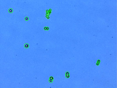
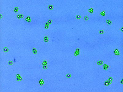
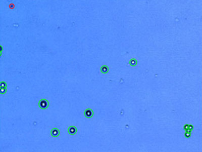
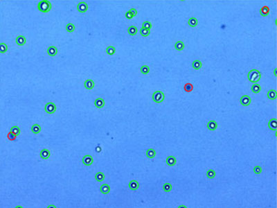
MEF: Fibroblastos embrionarios de roedor
 | |
Protocolo
|
Resultados
|
|---|
Reducción de ruido: 7
Redondez: 60%
Tamaño celular mín. (μm): 3
Tamaño celular máx. (μm): 60
Nivel de desagrupamiento: Ninguno
|
Concentración de células totales (células/ml): 1,03 x 10⁶
Concentración de células vivas (células/ml): 9,37 x 10⁵
Concentración de células muertas (células/ml): 9,72 x 10⁴
Viabilidad (%): 90,6
Tamaño celular promedio (μm): 19,6
|
|
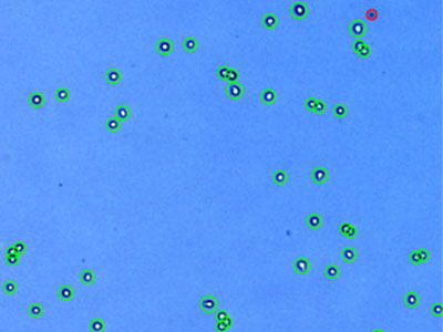
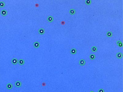
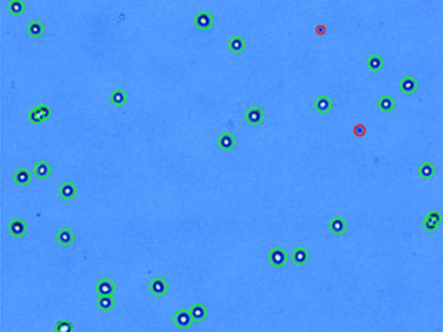
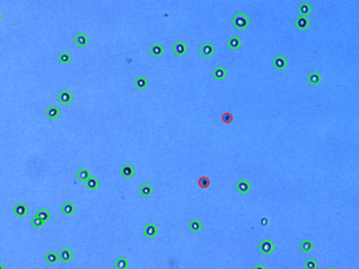
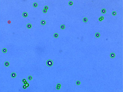
A549: Carcinoma de pulmón humano
 | |
Protocolo
|
Resultados
|
|---|
Reducción de ruido: 5
Redondez: 60%
Tamaño celular mín. (μm): 3
Tamaño celular máx. (μm): 60
Nivel de desagrupamiento: Medio
|
Concentración de células totales (células/ml): 1,09 x 10⁶
Concentración de células vivas (células/ml): 1,07 x 10⁶
Concentración de células muertas (células/ml): 1,78 x 10⁴
Viabilidad (%): 98,4
Tamaño celular promedio (μm): 17,5
|
|
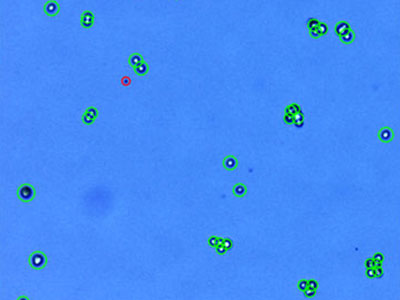
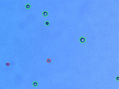
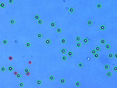
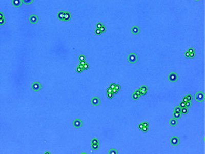
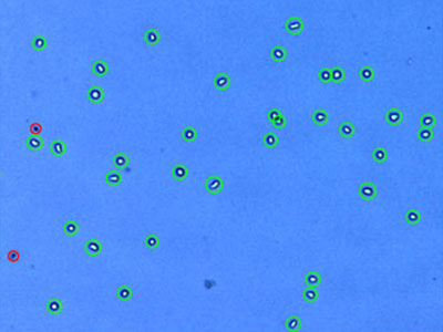
AsPC-1: Adenocarcinoma pancreático humano
 | |
Protocolo
|
Resultados
|
|---|
Reducción de ruido: 5
Redondez: 60%
Tamaño celular mín. (μm): 3
Tamaño celular máx. (μm): 60
Nivel de desagrupamiento: Alto
|
Concentración de células totales (células/ml): 7,51 x 10⁵
Concentración de células vivas (células/ml): 7,19 x 10⁵
Concentración de células muertas (células/ml): 3,17 x 10⁴
Viabilidad (%): 95,8
Tamaño celular promedio (μm): 10,7
|
|
C6: Glioma de rata
 | |
Protocolo
|
Resultados
|
|---|
Reducción de ruido: 5
Redondez: 60%
Tamaño celular mín. (μm): 3
Tamaño celular máx. (μm): 60
Nivel de desagrupamiento: Medio
|
Concentración de células totales (células/ml): 1,31 x 10⁶
Concentración de células vivas (células/ml): 1,22 x 10⁶
Concentración de células muertas (células/ml): 8,91 x 10⁴
Viabilidad (%): 93,2
Tamaño celular promedio (μm): 12,1
|
|
Capan-2:Adenocarcinoma pancreático humano
 | |
Protocolo
|
Resultados
|
|---|
Reducción de ruido: 5
Redondez: 60%
Tamaño celular mín. (μm): 3
Tamaño celular máx. (μm): 60
Nivel de desagrupamiento: Medio
|
Concentración de células totales (células/ml): 1,04 x 10⁶
Concentración de células vivas (células/ml): 1,00 x 10⁶
Concentración de células muertas (células/ml): 3,61 x 10⁴
Viabilidad (%): 96,5
Tamaño celular promedio (μm): 13,4
|
|
DU 145: Carcinoma prostático humano
 | |
Protocolo
|
Resultados
|
|---|
Reducción de ruido: 5
Redondez: 60%
Tamaño celular mín. (μm): 3
Tamaño celular máx. (μm): 60
Nivel de desagrupamiento: Medio
|
Concentración de células totales (células/ml): 1,86 x 10⁶
Concentración de células vivas (células/ml): 1,80 x 10⁶
Concentración de células muertas (células/ml): 5,34 x 10⁴
Viabilidad (%): 97,1
Tamaño celular promedio (μm): 14,3
|
|
293-GFP: HEK293 expresando GFP de forma estable
 | |
Protocolo
|
Resultados
|
|---|
Reducción de ruido: 5
Redondez: 60%
Tamaño celular mín. (μm): 3
Tamaño celular máx. (μm): 60
Nivel de desagrupamiento: Medio
|
Concentración de células totales (células/ml): 1,84 x 10⁶
Concentración de células vivas (células/ml): 1,82 x 10⁶
Concentración de células muertas (células/ml): 2,23 x 10⁴
Viabilidad (%): 98,8
Tamaño celular promedio (μm): 11,3
|
|
H1299: Carcinoma de pulmón no microcítico humano
 | |
Protocolo
|
Resultados
|
|---|
Reducción de ruido: 5
Redondez: 60%
Tamaño celular mín. (μm): 3
Tamaño celular máx. (μm): 60
Nivel de desagrupamiento: Medio
|
Concentración de células totales (células/ml): 4,14 x 10⁵
Concentración de células vivas (células/ml): 3,52 x 10⁵
Concentración de células muertas (células/ml): 6,23 x 10⁴
Viabilidad (%): 84,9
Tamaño celular promedio (μm): 19,5
|
|
H4:Neuroglioma humano
 | |
Protocolo
|
Resultados
|
|---|
Reducción de ruido: 5
Redondez: 60%
Tamaño celular mín. (μm): 3
Tamaño celular máx. (μm): 60
Nivel de desagrupamiento: Medio
|
Concentración de células totales (células/ml): 2,08 x 10⁵
Concentración de células vivas (células/ml): 1,44 x 10⁵
Concentración de células muertas (células/ml): 6,32 x 10⁴
Viabilidad (%): 69,6%
Tamaño celular promedio (μm): 12,8
|
|
HCT116: Carcinoma colorrectal humano
 | |
Protocolo
|
Resultados
|
|---|
Reducción de ruido: 5
Redondez: 60%
Tamaño celular mín. (μm): 3
Tamaño celular máx. (μm): 60
Nivel de desagrupamiento: Medio
|
Concentración de células totales (células/ml): 1,34 x 10⁶
Concentración de células vivas (células/ml): 1,32 x 10⁶
Concentración de células muertas (células/ml): 1,81 x 10⁴
Viabilidad (%): 98,7
Tamaño celular promedio (μm): 12,2
|
|
HeLa: Carcinoma cervical humano*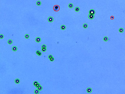 | |
Protocolo
|
Resultados
|
|---|
Reducción de ruido: 5
Redondez: 60%
Tamaño celular mín. (μm): 3
Tamaño celular máx. (μm): 60
Nivel de desagrupamiento: Medio
|
Concentración de células totales (células/ml): 8,98 x 10⁵
Concentración de células vivas (células/ml): 8,11 x 10⁵
Concentración de células muertas (células/ml): 8,69 x 10⁴
Viabilidad (%): 90,3
Tamaño celular promedio (μm): 17,6
|
|
Hep3B: Carcinoma hepatocelular humano
 | |
Protocolo
|
Resultados
|
|---|
Reducción de ruido: 5
Redondez: 60%
Tamaño celular mín. (μm): 3
Tamaño celular máx. (μm): 60
Nivel de desagrupamiento: Medio
|
Concentración de células totales (células/ml): 1,54 x 10⁶
Concentración de células vivas (células/ml): 1,49 x 10⁶
Concentración de células muertas (células/ml): 5,87 x 10⁴
Viabilidad (%): 96,2
Tamaño celular promedio (μm): 14,9
|
|
HL-60:Leucemia promielocítica aguda humana
 | |
Protocolo
|
Resultados
|
|---|
Reducción de ruido: 5
Redondez: 60%
Tamaño celular mín. (μm): 3
Tamaño celular máx. (μm): 60
Nivel de desagrupamiento: Medio
|
Concentración de células totales (células/ml): 1,57 x 10⁶
Concentración de células vivas (células/ml): 1,45 x 10⁶
Concentración de células muertas (células/ml): 1,23 x 10⁵
Viabilidad (%): 92,2
Tamaño celular promedio (μm): 14,9
|
|
Hs 578T:Carcinoma mamario humano
 | |
Protocolo
|
Resultados
|
|---|
Reducción de ruido: 5
Redondez: 60%
Tamaño celular mín. (μm): 3
Tamaño celular máx. (μm): 60
Nivel de desagrupamiento: Medio
|
Concentración de células totales (células/ml): 1,13 x 10⁶
Concentración de células vivas (células/ml): 8,72 x 10⁵
Concentración de células muertas (células/ml): 2,62 x 10⁵
Viabilidad (%): 76,9
Tamaño celular promedio (μm): 16,5
|
|
MCF7: Adenocarcinoma de mama humano
 | |
Protocolo
|
Resultados
|
|---|
Reducción de ruido: 5
Redondez: 60%
Tamaño celular mín. (μm): 3
Tamaño celular máx. (μm): 60
Nivel de desagrupamiento: Medio
|
Concentración de células totales (células/ml): 4,74 x 10⁵
Concentración de células vivas (células/ml): 4,43 x 10⁵
Concentración de células muertas (células/ml): 4,07 x 10⁴
Viabilidad (%): 91,4
Tamaño celular promedio (μm): 16,2
|
|
NIH3T3: Fibroblasto embrionario de ratón
 | |
Protocolo
|
Resultados
|
|---|
Reducción de ruido: 5
Redondez: 60%
Tamaño celular mín. (μm): 3
Tamaño celular máx. (μm): 60
Nivel de desagrupamiento: Medio
|
Concentración de células totales (células/ml): 1,52 x 10⁶
Concentración de células vivas (células/ml): 1,33 x 10⁶
Concentración de células muertas (células/ml): 1,96 x 10⁵
Viabilidad (%): 87,1
Tamaño celular promedio (μm): 14,1
|
|
PANC-1:Carcinoma ductal pancreático humano
 | |
Protocolo
|
Resultados
|
|---|
Reducción de ruido: 5
Redondez: 60%
Tamaño celular mín. (μm): 3
Tamaño celular máx. (μm): 60
Nivel de desagrupamiento: Alto
|
Concentración de células totales (células/ml): 1,23 x 10⁶
Concentración de células vivas (células/ml): 1,21 x 10⁵
Concentración de células muertas (células/ml): 2,26 x 10⁴
Viabilidad (%): 98,2
Tamaño celular promedio (μm): 15,3
|
|
PC-3: Adenocarcinoma prostático humano
 | |
Protocolo
|
Resultados
|
|---|
Reducción de ruido: 5
Redondez: 60%
Tamaño celular mín. (μm): 3
Tamaño celular máx. (μm): 60
Nivel de desagrupamiento: Medio
|
Concentración de células totales (células/ml): 1,36 x 10⁶
Concentración de células vivas (células/ml): 1,33 x 10⁶
Concentración de células muertas (células/ml): 3,16 x 10⁴
Viabilidad (%): 97,7
Tamaño celular promedio (μm): 17,2
|
|
STO: Fibroblasto embrionario de ratón
 | |
Protocolo
|
Resultados
|
|---|
Reducción de ruido: 5
Redondez: 0%
Tamaño celular mín. (μm): 3
Tamaño celular máx. (μm): 60
Nivel de desagrupamiento: Medio
|
Concentración de células totales (células/ml): 1,78 x 10⁶
Concentración de células vivas (células/ml): 1,50 x 10⁶
Concentración de células muertas (células/ml): 2,81 x 10⁵
Viabilidad (%): 84,2
Tamaño celular promedio (μm): 13,1
|
|
U-2 OS: Osteosarcoma humano
 | |
Protocolo
|
Resultados
|
|---|
Reducción de ruido: 5
Redondez: 60%
Tamaño celular mín. (μm): 3
Tamaño celular máx. (μm): 60
Nivel de desagrupamiento: Medio
|
Concentración de células totales (células/ml): 7,66 x 10⁵
Concentración de células vivas (células/ml): 7,21 x 10⁵
Concentración de células muertas (células/ml): 4,45 x 10⁴
Viabilidad (%): 94,2
Tamaño celular promedio (μm): 16,0
|
|
U937: Linfoma humano
 | |
Protocolo
|
Resultados
|
|---|
Reducción de ruido: 5
Redondez: 40%
Tamaño celular mín. (μm): 3
Tamaño celular máx. (μm): 60
Nivel de desagrupamiento: Medio
|
Concentración de células totales (células/ml): 1,22 x 10⁶
Concentración de células vivas (células/ml): 1,12 x 10⁶
Concentración de células muertas (células/ml): 1,02 x 10⁵
Viabilidad (%): 91,6
Tamaño celular promedio (μm): 13,4
|
|
* Si bien estas líneas celulares forman parte de las más importantes para la investigación médica, es imperativo reconocer la contribución de Henrietta Lacks a la ciencia que se produjo sin su consentimiento. Esta injusticia, a pesar de haber dado lugar a descubrimientos clave en inmunología, enfermedades infecciosas y cáncer, también ha generado importantes debates sobre la privacidad, la ética y el consentimiento en la medicina.
Para obtener más información sobre la vida de Henrietta Lacks y su contribución a la medicina moderna, haga clic aquí.
http://henriettalacksfoundation.org/
|